You have no items in your shopping cart.
Description
Research Area
Cardiovascular Research
Images & Validation
−Item 1 of 4
| Tested Applications | FC |
|---|---|
| Reactivity | Human, Mouse |
| Application Notes |
Key Properties
−| Antibody Type | Primary Antibody |
|---|---|
| Clonality | Monoclonal |
| Isotype | Mouse IgG1 kappa |
| Clone No. | mN1A |
| Immunogen | GST fusion protein containing cdc10-NCR region of mouse Notch1 |
| Target | Notch1 |
| Purification | Purified antibody is conjugated with activated allophycocyanin (APC) under optimum conditions and unconjugated antibody and free fluorochrome are removed by size-exclusion chromatography. |
| Conjugation | APC |
Storage & Handling
−| Storage | Store at 2-8°C. Protect from prolonged exposure to light. Do not freeze. |
|---|---|
| Buffer/Preservatives | Stabilizing phosphate buffered saline (PBS), pH 7.4, 15 mM sodium azide |
| Concentration | 0.1 mg/ml |
| Expiration Date | 12 months from date of receipt. |
| Disclaimer | For research use only |
Alternative Names
−AOS5, TAN1, hN1, AOVD1
Similar Products
−
- Item 1 of 1
Activated Notch1 Rabbit Polyclonal Antibody (APC) [orb999156]
IF
Bovine, Canine, Guinea pig, Human, Porcine, Rabbit, Rat
Mouse
Rabbit
Polyclonal
APC
100 μl
Quality Guarantee
Explore bioreagents carefree to elevate your research. All our products are rigorously tested for performance. If a product does not perform as described on its datasheet, our scientific support team will provide expert troubleshooting, a prompt replacement, or a refund. For full details, please see our Terms & Conditions and Buying Guide. Contact us at [email protected].
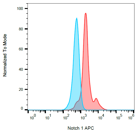
Separation of MOLT-4 cell line (red) from Leukocytes(blue) in flow cytometry analysis (intracellular staining) stained using anti-human Notch1 (mN1A) APC antibody (concentration in sample 3 µg/ml).
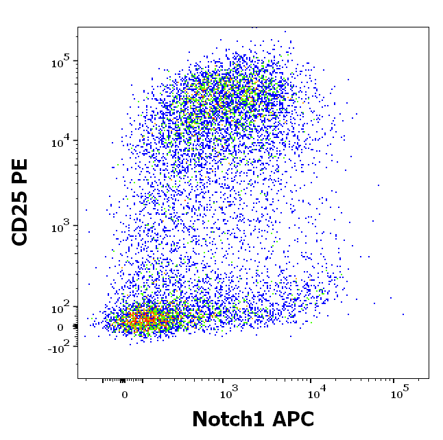
Flow cytometry multicolor surface staining pattern of human PHA stimulated CD3 positive lymphocytes using anti-human CD25 (MEM-181) PE antibody (20 µl reagent / 100 µl of sample) and intracellular staining of human lymphocytes using anti-Notch1 (mN1A) APC antibody (concentration in sample 3 µg/ml).
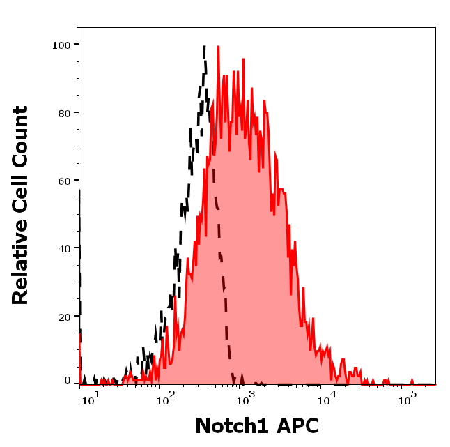
Separation of CD3 positive CD25 positive cells stained using anti-Notch1 (mN1A) APC antibody (concentration in sample 3 µg/ml, red-filled) from CD3 positive CD25 positive cells stained using mouse IgG1 isotype control (MOPC-21) APC antibody (concentration in sample 3 µg/ml, same as Notch1 APC concentration, black-dashed) in flow cytometry analysis (intracellular staining) of PHA stimulated human peripheral whole blood.
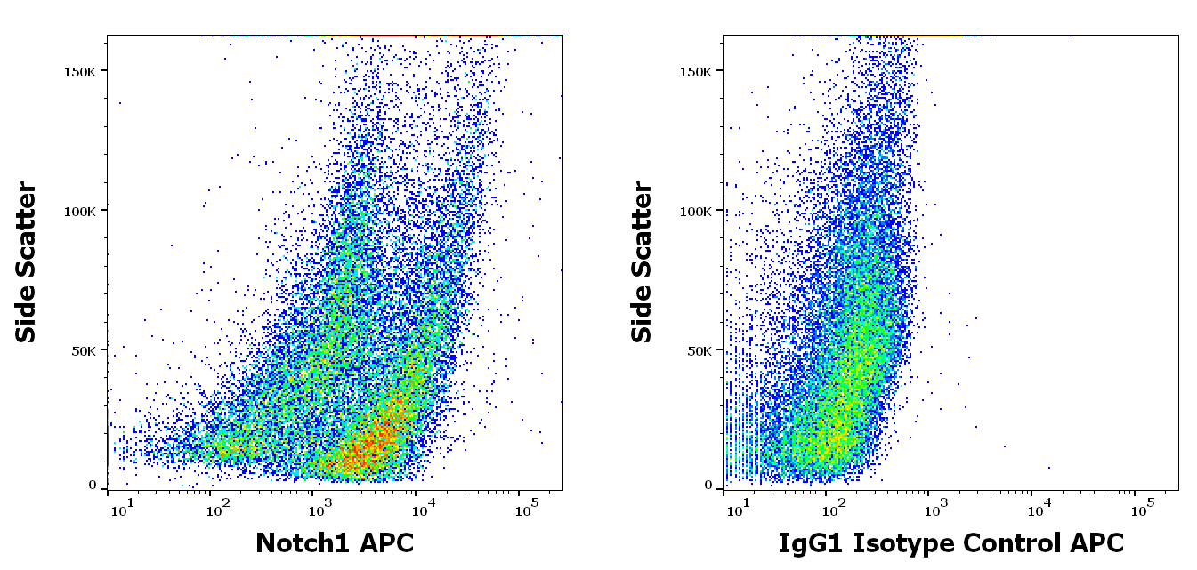
Flow cytometry intracellular staining patterns of PHA stimulated human peripheral whole blood stained using anti-Notch1 (mN1A) PE antibody (concentration in sample 3 µg/ml, left) or mouse IgG1 isotype control (MOPC-21) PE antibody (concentration in sample 3 µg/ml, same as Notch1 PE concentration, right).
Documents Download
Datasheet
Product Information
Request a Document
Notch1 Antibody (APC) (orb154447)

- 0.0
Based on 0 reviews
Participating in our Biorbyt product reviews program enables you to support fellow scientists by sharing your firsthand experience with our products.
Login to Submit a ReviewAvailable Sizes
Select a size below
Free Secondary Antibody (20 ul)0/0
Please add an antibody product to your cart first.