You have no items in your shopping cart.
Featured
Description
Research Area
Cancer Biology, Cell Biology, Neuroscience
Images & Validation
−Item 1 of 3
| Tested Applications | ELISA, ICC, IF, IHC, WB |
|---|---|
| Dilution Range | WB (1:1000) |
| Reactivity | Human, Mouse, Rat |
| Application Notes |
Key Properties
−| Host | Mouse |
|---|---|
| Clonality | Monoclonal |
| Isotype | IgG2a |
| Clone No. | J23 |
| Immunogen | Partial Human Recombinant NOS1 Protein |
| Target | NOS1 |
| Molecular Weight | 160 kDa |
| Purification | Protein A Purified |
| Conjugation | APC |
Storage & Handling
−| Storage | Conjugated antibodies should be stored according to the product label |
|---|---|
| Buffer/Preservatives | 95.46mM Phosphate, 2.48mM MES and 2mM EDTA |
| Concentration | 1mg/ml |
| Expiration Date | 12 months from date of receipt. |
| Disclaimer | For research use only |
Alternative Names
−BNOS, Constitutive NOS, IHPS 1, IHPS1, N-NOS, NC-NOS, neuronal Nitric Oxide Synthase, Neuronal NOS, Nitric oxide synthase neuronal included, Nitric oxide synthase 1 (neuronal), Nitric oxide synthase 1, NNOS, NO, NOS 1, NOS, NOS type I, NOS-I, NOS1, NOS1_HUMAN
Similar Products
−
Phospho-nNOS (Ser1417) Rabbit Polyclonal Antibody (APC) [orb1001759]
IF
Human, Mouse, Xenopus, Zebrafish
Rat
Rabbit
Polyclonal
APC
100 μlPhospho-nNOS (Ser847) Rabbit Polyclonal Antibody (APC) [orb1001760]
IF
Equine, Gallus, Mouse, Porcine, Rabbit, Rat, Xenopus
Human
Rabbit
Polyclonal
APC
100 μlNNOS Rabbit Polyclonal Antibody (APC) [orb1005707]
IF
Human, Mouse, Rabbit
Rat
Rabbit
Polyclonal
APC
100 μlNNOS Rabbit Polyclonal Antibody (APC) [orb996530]
IF
Bovine, Canine, Gallus, Human, Porcine, Rat, Sheep
Mouse
Rabbit
Polyclonal
APC
100 μlPhospho-nNOS (Ser847) Rabbit Polyclonal Antibody (APC-Cy7) [orb2470841]
IF
Equine, Gallus, Mouse, Porcine, Rabbit, Rat, Xenopus
Human
Rabbit
Polyclonal
APC/Cy7
100 μl
Quality Guarantee
Explore bioreagents carefree to elevate your research. All our products are rigorously tested for performance. If a product does not perform as described on its datasheet, our scientific support team will provide expert troubleshooting, a prompt replacement, or a refund. For full details, please see our Terms & Conditions and Buying Guide. Contact us at [email protected].
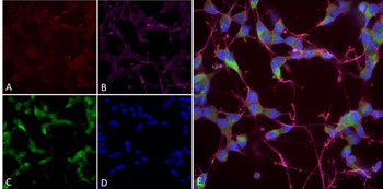
Immunocytochemistry/Immunofluorescence analysis using Mouse Anti-NOS1/ nNos Monoclonal Antibody, Clone J23. Tissue: Differentiated SH-SY5Y. Species: Human. Primary Antibody: Mouse Anti-NOS1/ nNos Monoclonal Antibody at 1:250. Secondary Antibody: AlexaFluor 488. Counterstain: phalloidin (Alexa 647, red), beta tubulin (Anti-beta III Tubulin Ab, Alexa 555, magenta) Hoechst (blue). (A) Phalloidin (B) Anti-beta III Tubulin Ab. (C) NOS1/nNOS Antibody. (D) Hoechst (E) Composite.
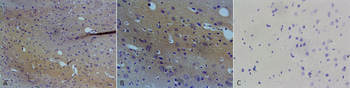
Immunohistochemistry analysis using Mouse Anti-NOS1/ nNOS Monoclonal Antibody, Clone J23. Tissue: brain tissue. Species: Rat. Primary Antibody: Mouse Anti-NOS1/ nNOS Monoclonal Antibody at 1:100. Secondary Antibody: Goat anti-mouse IgG1: HRP at 1:100. Counterstain: DAPI at 1 ug/ml. A) 10X magnification. B) 20X magnification. C) Negative control mouse IgG (10 ug/ml), 20X magnification.
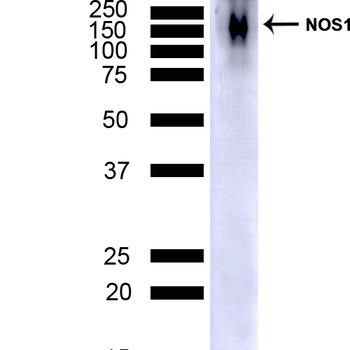
Western Blot analysis of Mouse Cerebellum showing detection of NOS1/ nNos protein using Mouse Anti-NOS1/ nNos Monoclonal Antibody, Clone J23. Lane 1: MW ladder. Lane 2: Mouse Brain Cerebellum (7.5 ug). Load: 7.5 ug. Block: 5% Skim Milk powder in TBST. Primary Antibody: Mouse Anti-NOS1/ nNos Monoclonal Antibody at 1:1000 for 2 hours at RT with shaking. Secondary Antibody: Goat anti-mouse IgG:HRP at 1:4000 for 1 hour at RT with shaking. Color Development: Chemiluminescent for HRP (Moss) for 5 min in RT.
Quick Database Links
UniProt Details
− No UniProt data available
NCBI Gene Details
− No NCBI Gene data available
NCBI Reference Sequences
−Associated Accession Numbers
Curated reference sequences for the gene transcript and protein product| Protein | NP_000611.1 |
|---|
Documents Download
Datasheet
Product Information
Request a Document
Protocol Information
WB
Western Blot (IB, immunoblot)
IHC
Immunohistochemistry
IF
Immunofluorescence
ICC
Immunocytochemistry
ELISA
Enzyme-linked Immunosorbent Assay (EIA)
NOS1 Antibody (APC) (orb699416)

- 0.0
Based on 0 reviews
Participating in our Biorbyt product reviews program enables you to support fellow scientists by sharing your firsthand experience with our products.
Login to Submit a ReviewAvailable Sizes
Select a size below
Choose Conjugation or Carrier Free Version
Free Secondary Antibody (20 ul)0/0
Please add an antibody product to your cart first.