You have no items in your shopping cart.
Description
Research Area
Cancer Biology, Neuroscience, Pharmacology & Drug Discovery
Images & Validation
−Item 1 of 4
| Tested Applications | ICC, IEM, IF, IHC, WB |
|---|---|
| Dilution Range | WB (1:1000), IHC (1:200) |
| Reactivity | Canine, Human, Mouse, Rat |
| Application Notes |
Key Properties
−| Host | Rabbit |
|---|---|
| Clonality | Polyclonal |
| Immunogen | Produced against a synthetic peptide mapping to a segment of rat NCC (amino acids 74-95), N-terminal |
| Target | NCC |
| Molecular Weight | 160kDa |
| Purification | Affinity Purified |
| Conjugation | APC |
Storage & Handling
−| Storage | Conjugated antibodies should be stored according to the product label |
|---|---|
| Buffer/Preservatives | 95.46mM Phosphate, 2.48mM MES and 2mM EDTA |
| Concentration | 1 mg/ml |
| Expiration Date | 12 months from date of receipt. |
| Disclaimer | For research use only |
Alternative Names
−NCC, SLC12A3, SCYL1, CKb10, MCP-4, MGC17134, NCC-1, NCC1, SCYA13, CK-beta-10, Monocyte chemoattractant protein 4, Monocyte chemotactic protein 4, New CC chemokine 1, Small inducible cytokine A13, Small inducible cytokine subfamily A (Cys-Cys) member 13, Chemokine (C-C)
Similar Products
−
CCL14/NCC-2 Mouse Monoclonal Antibody (APC) [orb2972624]
ELISA, FC
Human
Mouse
Monoclonal
APC
50 T, 100 TCCL13 Rabbit Polyclonal Antibody (APC) [orb1004595]
ICC, IF
Bovine, Canine, Equine, Human, Rabbit
Rabbit
Polyclonal
APC
100 μl
Quality Guarantee
Explore bioreagents carefree to elevate your research. All our products are rigorously tested for performance. If a product does not perform as described on its datasheet, our scientific support team will provide expert troubleshooting, a prompt replacement, or a refund. For full details, please see our Terms & Conditions and Buying Guide. Contact us at [email protected].
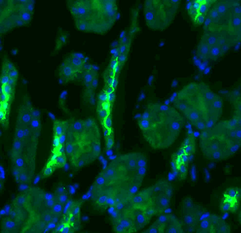
Immunohistochemistry analysis using Rabbit Anti-NCC Polyclonal Antibody. Tissue: kidney tissue. Species: Rat. Primary Antibody: Rabbit Anti-NCC Polyclonal Antibody at 1:200. Secondary Antibody: FITC Goat Anti-Rabbit (green).
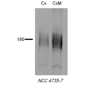
Western blot analysis of Rat tissue lysates showing detection of NCC protein using Rabbit Anti-NCC Polyclonal Antibody. Primary Antibody: Rabbit Anti-NCC Polyclonal Antibody at 1:1000.
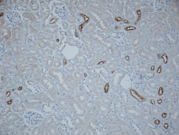
Immunohistochemistry analysis using Rabbit Anti-NCC Polyclonal Antibody. Tissue: kidney tissue. Species: Mouse. Fixation: paraformaldehyde-fixed paraffin-embedded. Primary Antibody: Rabbit Anti-NCC Polyclonal Antibody at 1:500 for Overnight at 4°C. Secondary Antibody: Biotinylated Goat Anti-Rabbit IgG at 1:500 for 30 min at RT.

Immunohistochemistry analysis using Rabbit Anti-NCC Polyclonal Antibody. Tissue: kidney tissue. Species: Mouse. Fixation: paraformaldehyde-fixed paraffin-embedded. Primary Antibody: Rabbit Anti-NCC Polyclonal Antibody at 1:1000 for Overnight at 4°C. Secondary Antibody: Biotinylated Goat Anti-Rabbit IgG at 1:501 for 30 min at RT.
Quick Database Links
Documents Download
Datasheet
Product Information
Request a Document
Protocol Information
WB
Western Blot (IB, immunoblot)
IHC
Immunohistochemistry
IF
Immunofluorescence
ICC
Immunocytochemistry
NCC Antibody (APC) (orb152699)

- 0.0
Based on 0 reviews
Participating in our Biorbyt product reviews program enables you to support fellow scientists by sharing your firsthand experience with our products.
Login to Submit a ReviewAvailable Sizes
Select a size below
Choose Conjugation or Carrier Free Version
Free Secondary Antibody (20 ul)0/0
Please add an antibody product to your cart first.