You have no items in your shopping cart.
Featured
Description
Research Area
Cell Biology
Images & Validation
−Item 1 of 4
| Tested Applications | IF, IHC-P, WB |
|---|---|
| Dilution Range | IF - 1:25, WB - 1:2000, IHC-P - 1:25 |
| Reactivity | Human, Mouse |
Key Properties
−| Host | Rabbit |
|---|---|
| Clonality | Polyclonal |
| Isotype | Rabbit IgG |
| Immunogen | This Sirt1 antibody is generated from a rabbit immunized with a KLH conjugated synthetic peptide between 566-601 amino acids from the C-terminal region of human Sirt1. |
| Target | Sirt1 |
| Molecular Weight | 80372 |
| Conjugation | Unconjugated |
Storage & Handling
−| Storage | Maintain refrigerated at 2-8°C for up to 2 weeks. For long term storage store at -20°C in small aliquots to prevent freeze-thaw cycles |
|---|---|
| Form/Appearance | Purified polyclonal antibody supplied in PBS with 0.09% (W/V) sodium azide. This antibody is purified through a protein A column, followed by peptide affinity purification. |
| Expiration Date | 12 months from date of receipt. |
| Disclaimer | For research use only |
Alternative Names
−anti NAD-dependent protein deacetylase sirtuin-1 antibody, anti 351- antibody, anti Regulatory protein SIR2 homolog 1 antibody, anti SIR2-like protein 1 antibody, anti SIR2alpha antibody, anti Sir2 antibody, anti mSIR2a antibody, anti SirtT1 75 kDa fragment antibody, anti 75SirT1 antibody, anti Sirt1 antibody, anti Sir2l1 antibody
Similar Products
−- Item 1 of 3
- Item 1 of 1

SIRT1 Rabbit Polyclonal Antibody [orb2988741]
WB
Human, Mouse
Rabbit
Polyclonal
Unconjugated
200 μl, 100 μl, 50 μl, 30 μl
Quality Guarantee
Explore bioreagents carefree to elevate your research. All our products are rigorously tested for performance. If a product does not perform as described on its datasheet, our scientific support team will provide expert troubleshooting, a prompt replacement, or a refund. For full details, please see our Terms & Conditions and Buying Guide. Contact us at [email protected].
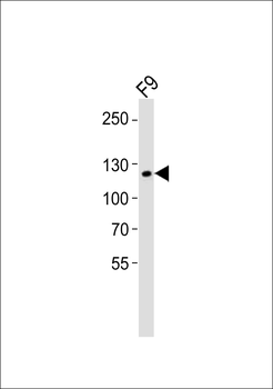
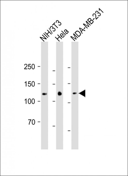
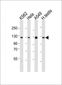
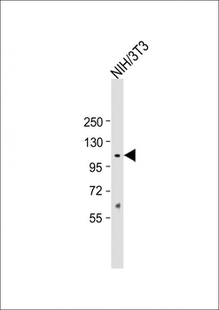
Western blot analysis of lysate from mouse F9 cell line, using Mouse Sirt1 Antibody (C-term). Diluted at 1:1000. A goat anti-rabbit IgG H&L (HRP) at 1:10000 dilution was used as the secondary antibody. Lysate at 20 ug.
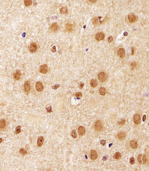
Immunohistochemical analysis of paraffin-embedded M. brain section using Mouse Sirt1 Antibody (C-term). Diluted at 1:25 dilution. A undiluted biotinylated goat polyvalent antibody was used as the secondary, followed by DAB staining.
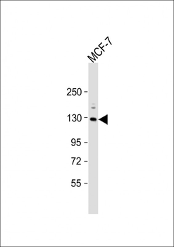
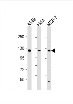
Anti-Sirt1 Antibody (C-term) at 1:2000 dilution + MCF-7 whole cell lysate. Lysates/proteins at 20 µg per lane. Secondary Goat Anti-Rabbit IgG, (H+L), Peroxidase conjugated at 1/10000 dilution. Predicted band size: 80 kDa. Blocking/Dilution buffer: 5% NFDM/TBST.
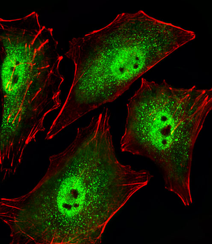
Fluorescent image of A549 cells stained with Mouse Sirt1 Antibody (C-term). Diluted at 1:25 dilution. An Alexa Fluor 488-conjugated goat anti-rabbit lgG at 1:400 dilution was used as the secondary antibody (green). Cytoplasmic actin was counterstained with Alexa Fluor 555 conjugated with Phalloidin (red).
Quick Database Links
Gene Symbol
Sirt1
UniProt
UniProt Details
− No UniProt data available
Documents Download
Datasheet
Product Information
Request a Document
Protocol Information
WB
Western Blot (IB, immunoblot)
IHC-P
Immunohistochemistry Paraffin
IF
Immunofluorescence
D'Onofrio, Nunzia et al. Synergistic Effect of Dietary Betaines on SIRT1-Mediated Apoptosis in Human Oral Squamous Cell Carcinoma Cal 27 Cancers (Basel), 12, E2468 (2020)
Sirt1 Antibody (C-term) (orb306144)

- 0.0
Based on 0 reviews
Participating in our Biorbyt product reviews program enables you to support fellow scientists by sharing your firsthand experience with our products.
Login to Submit a ReviewAvailable Sizes
Select a size below
Choose Conjugation or Carrier Free Version
Free Secondary Antibody (20 ul)0/0
Please add an antibody product to your cart first.