You have no items in your shopping cart.
Description
Research Area
Cancer Biology
Images & Validation
−Item 1 of 3
| Tested Applications | FC, WB |
|---|---|
| Dilution Range | WB - 1:1000, FC - 1:25 |
| Reactivity | Mouse |
Key Properties
−| Host | Rabbit |
|---|---|
| Clonality | Polyclonal |
| Isotype | Rabbit IgG |
| Immunogen | This Mouse Klf4 antibody is generated from a rabbit immunized with a KLH conjugated synthetic peptide between 321-354 amino acids from the Central region of mouse Klf4. |
| Target | Klf4 |
| Molecular Weight | 51880 Da |
| Conjugation | Unconjugated |
Storage & Handling
−| Storage | Maintain refrigerated at 2-8°C for up to 2 weeks. For long term storage store at -20°C in small aliquots to prevent freeze-thaw cycles |
|---|---|
| Form/Appearance | Purified polyclonal antibody supplied in PBS with 0.09% (W/V) sodium azide. This antibody is purified through a protein A column, followed by peptide affinity purification. |
| Expiration Date | 12 months from date of receipt. |
| Disclaimer | For research use only |
Alternative Names
−Krueppel-like factor 4, Epithelial zinc finger protein EZF, Gut-enriched krueppel-like factor, Klf4, Ezf, Gklf, Zie
Similar Products
−- Item 1 of 2
Mouse Klf4 Antibody (Center) [orb1788068]
FC, WB
Mouse
Rabbit
Polyclonal
Unconjugated

Quality Guarantee
Explore bioreagents carefree to elevate your research. All our products are rigorously tested for performance. If a product does not perform as described on its datasheet, our scientific support team will provide expert troubleshooting, a prompt replacement, or a refund. For full details, please see our Terms & Conditions and Buying Guide. Contact us at [email protected].
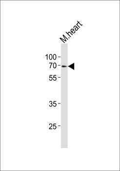
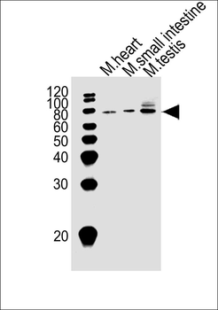
Western blot analysis of lysate from mouse heart tissue lysate, using Klf4 Antibody (Center). Diluted at 1:1000. A goat anti-rabbit IgG H&L (HRP) at 1:10000 dilution was used as the secondary antibody. Lysate at 20 ug.
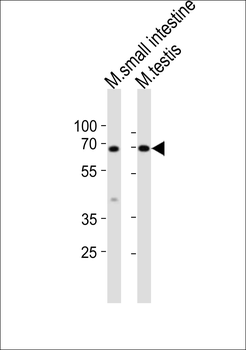
Western blot analysis of lysates from mouse small intestine, mouse testis tissue lysate (from left to right), using Klf4 Antibody (Center). Diluted at 1:1000 at each lane. A goat anti-rabbit IgG H&L (HRP) at 1:10000 dilution was used as the secondary antibody. Lysates at 20 ug per lane.
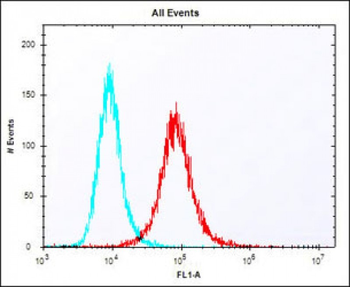
Overlay histogram showing NIH/3T3 cells stained (red line). The cells were fixed with 2% paraformaldehyde (10 min) and then permeabilized with 90% methanol for 10 min. The cells were then icubated in 2% bovine serum albumin to block non-specific protein-protein interactions followed by the antibody (1:25 dilution) for 60 min at 37°C. The secondary antibody used was Alexa Fluor 488 goat anti-rabbit lgG (H+L) (1583138) at 1/400 dilution for 40 min at 37°C. Isotype control antibody (blue line) was rabbit IgG1 (1 μg/1x10^6 cells) used under the same conditions. Acquisition of > 10000 events was performed.
Quick Database Links
Gene Symbol
Klf4
UniProt
UniProt Details
− No UniProt data available
Documents Download
Datasheet
Product Information
Request a Document
Protocol Information
WB
Western Blot (IB, immunoblot)
FC
Flow Cytometry
Mouse Klf4 Antibody (Center) (orb1926632)

- 0.0
Based on 0 reviews
Participating in our Biorbyt product reviews program enables you to support fellow scientists by sharing your firsthand experience with our products.
Login to Submit a ReviewAvailable Sizes
Select a size below
Choose Conjugation or Carrier Free Version
Free Secondary Antibody (20 ul)0/0
Please add an antibody product to your cart first.