You have no items in your shopping cart.
Description
Research Area
Immunology & Inflammation
Images & Validation
−Item 1 of 4
| Tested Applications | FC, IHC-P, WB |
|---|---|
| Dilution Range | WB - 1:1000, IHC-P - 1:25, FC - 1:25 |
| Reactivity | Mouse |
| Predicted Reactivity | Rat |
Key Properties
−| Host | Rabbit |
|---|---|
| Clonality | Polyclonal |
| Isotype | Rabbit IgG |
| Immunogen | This mouse Epcam antibody is generated from a rabbit immunized with a KLH conjugated synthetic peptide between 302-335 amino acids from the C-terminal region of mouse Epcam. |
| Target | Epcam |
| Molecular Weight | 35019 Da |
| Conjugation | Unconjugated |
Storage & Handling
−| Storage | Maintain refrigerated at 2-8°C for up to 2 weeks. For long term storage store at -20°C in small aliquots to prevent freeze-thaw cycles |
|---|---|
| Form/Appearance | Purified polyclonal antibody supplied in PBS with 0.09% (W/V) sodium azide. This antibody is purified through a protein A column, followed by peptide affinity purification. |
| Expiration Date | 12 months from date of receipt. |
| Disclaimer | For research use only |
Alternative Names
−Epithelial cell adhesion molecule, Ep-CAM, Epithelial glycoprotein 314, EGP314, mEGP314, Protein 289A, Tumor-associated calcium signal transducer 1, CD326, Epcam, Tacstd1
Similar Products
−- Item 1 of 3
(Mouse) Epcam Antibody (C-term) [orb1926423]
IHC-P, WB
Human, Mouse, Rat
Rabbit
Polyclonal
Unconjugated
50 μl, 100 μl - Item 1 of 2
(Mouse) Epcam Antibody (C-term) [orb1926698]
FC, WB
Rat
Mouse
Rabbit
Polyclonal
Unconjugated
50 μl, 100 μl - Item 1 of 2
(Mouse) Epcam Antibody (C-term) [orb1788010]
FC, WB
Human, Mouse
Rabbit
Polyclonal
Unconjugated

Quality Guarantee
Explore bioreagents carefree to elevate your research. All our products are rigorously tested for performance. If a product does not perform as described on its datasheet, our scientific support team will provide expert troubleshooting, a prompt replacement, or a refund. For full details, please see our Terms & Conditions and Buying Guide. Contact us at [email protected].
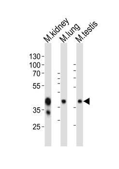
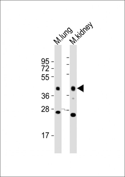
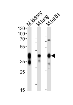
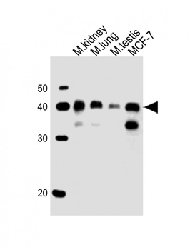
Western blot analysis of lysates from mouse kidney, mouse lung, mouse testis tissue lysate (from left to right), using Epcam Antibody (C-term). Diluted at 1:1000 at each lane. A goat anti-rabbit IgG H&L (HRP) at 1:10000 dilution was used as the secondary antibody. Lysates at 20 ug per lane.
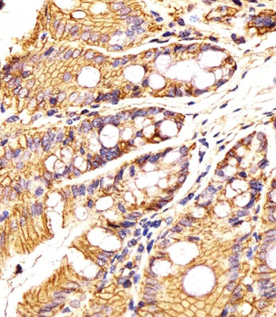
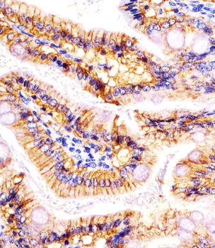
Staining Epcam in Mouse colon tissue sections by Immunohistochemistry (IHC-P - paraformaldehyde-fixed, paraffin-embedded sections). Tissue was fixed with formaldehyde and blocked with 3% BSA for 0.5 hour at room temperature; antigen retrieval was by heat mediation with a citrate buffer (pH6). Samples were incubated with primary antibody (1/25) for 1 hours at 37°C. A undiluted biotinylated goat polyvalent antibody was used as the secondary antibody.
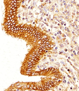
Staining Epcam in Human colorectal carcinoma tissue sections by Immunohistochemistry (IHC-P - paraformaldehyde-fixed, paraffin-embedded sections). Tissue was fixed with formaldehyde and blocked with 3% BSA for 0.5 hour at room temperature; antigen retrieval was by heat mediation with a citrate buffer (pH6). Samples were incubated with primary antibody (1/25) for 1 hours at 37°C. A undiluted biotinylated goat polyvalent antibody was used as the secondary antibody.
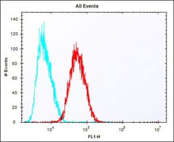
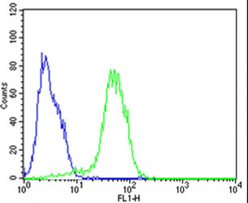
Overlay histogram showing HepG2 cells stained (red line). The cells were fixed with 2% paraformaldehyde (10 min) and then permeabilized with 90% methanol for 10 min. The cells were then icubated in 2% bovine serum albumin to block non-specific protein-protein interactions followed by the antibody (1:25 dilution) for 60 min at 37°C. The secondary antibody used was Alexa Fluor 488 goat anti-rabbit lgG (H+L) (1583138) at 1/400 dilution for 40 min at 37°C. Isotype control antibody (blue line) was rabbit IgG1 (1 μg/1x10^6 cells) used under the same conditions. Acquisition of > 10000 events was performed.
Quick Database Links
Gene Symbol
Epcam
UniProt
UniProt Details
− No UniProt data available
Documents Download
Datasheet
Product Information
Request a Document
Protocol Information
WB
Western Blot (IB, immunoblot)
IHC-P
Immunohistochemistry Paraffin
FC
Flow Cytometry
(Mouse) Epcam Antibody (C-term) (orb1926651)

- 0.0
Based on 0 reviews
Participating in our Biorbyt product reviews program enables you to support fellow scientists by sharing your firsthand experience with our products.
Login to Submit a ReviewAvailable Sizes
Select a size below
Choose Conjugation or Carrier Free Version
Free Secondary Antibody (20 ul)0/0
Please add an antibody product to your cart first.