You have no items in your shopping cart.
Featured
Description
Research Area
Immunology & Inflammation
Images & Validation
−Item 1 of 5
| Tested Applications | FC, IF, IHC, WB |
|---|---|
| Dilution Range | FC: Fixed in 4% formaldehyde and permeabilized with 90% methanol. 0.25 µl per 1 x 10^6 cells. Immunohistochemistry (IHC): 1:100 - 1:500. Epitope retrieval with citrate buffer pH 6.0 is recommended for FFPE tissue sections. IHC-IF: 1:100 - 1:500. Epitope retrieval with citrate buffer pH6.0 is recommended for FFPE tissue sections. Multiplex Immunofluorescence (mIF): 1:250. WB: 1:1000 |
| Reactivity | Human |
| Application Notes |
Key Properties
−| Antibody Type | Primary Antibody |
|---|---|
| Host | Mouse |
| Clonality | Monoclonal |
| Isotype | IgG2a |
| Clone No. | L26 |
| Immunogen | Human tonsil B cells |
| Target | CD20 |
| Purification | Purified |
| Conjugation | Unconjugated |
Customer Validated Data
− The data below is submitted by researchers worldwide. We provide this transparency to help you decide if this product works for your specific application or species, even if we haven't tested it ourselves.
Success by Application
Reactivity Distribution
Latest Experiments
1 ResultsDilution
Sample
Summary
IF
Human
-
Tissue microarrays (TMA) constructed from FFPE
Sample preparation involved deparaffinizing TMA sections...
Storage & Handling
−| Storage | 2 - 8°C |
|---|---|
| Form/Appearance | Liquid |
| Buffer/Preservatives | Phosphate Buffered Saline (PBS) pH 8.2 with 0.1% rAlbumin and 0.09% Sodium Azide |
| Concentration | 300 µg/ml |
| Expiration Date | 12 months from date of receipt. |
| Disclaimer | For research use only |
Alternative Names
−B1; B-lymphocyte antigen CD20; B-lymphocyte cell-surface antigen B1; B-lymphocyte surface antigen B1; Bp35; CD antigen CD20; CD20; CD20 antigen; CD20 receptor; CVID5; LEU-16; leukocyte surface antigen Leu-16; Membrane-spanning 4-domains subfamily A member 1; membrane-spanning 4-domains, subfamily A, member 1; MS4A2; S7
Similar Products
−- Item 1 of 8
Caspase 8 Mouse Monoclonal Antibody [orb500937]
WB
Rat
Human, Mouse
Mouse
Monoclonal
Unconjugated
50 μl, 100 μl, 200 μl, 200 μg - Item 1 of 7
Cytokeratin 20 Mouse Monoclonal Antibody [orb1816739]
IF, IHC-Fr, IHC-P
Mouse, Rat
Human, Mouse, Rat
Mouse
Monoclonal
Unconjugated
50 μl, 100 μl - Item 1 of 5
MS4A1 Antibody [orb1410317]
IHC, WB
Human, Mouse, Rat
Mouse
Monoclonal
Unconjugated
20 μg, 100 μg, 100 μg (without BSA and Azide) - Item 1 of 4
- Item 1 of 3


![Anti-CD20 [10F381 (rituximab)]](/images/pub/media/catalog/product/NewWebsite/35/orb396140_1.png)
![Anti-CD20 [10F381 (rituximab)]](/images/pub/media/catalog/product/NewWebsite/35/orb396140_2.png)
![Anti-CD20 [10F381 (rituximab)]](/images/pub/media/catalog/product/NewWebsite/35/orb396140_3.png)
Quality Guarantee
Explore bioreagents carefree to elevate your research. All our products are rigorously tested for performance. If a product does not perform as described on its datasheet, our scientific support team will provide expert troubleshooting, a prompt replacement, or a refund. For full details, please see our Terms & Conditions and Buying Guide. Contact us at [email protected].
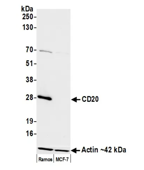
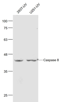
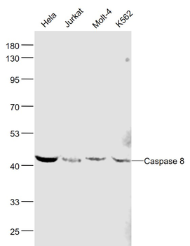
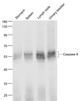
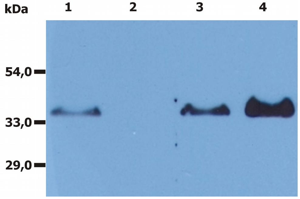
Detection of human CD20 by western blot. Samples: Whole cell lysate (25 µg) from Ramos and MCF-7 cells prepared using NETN lysis buffer. Antibody: Mouse anti-CD20 monoclonal antibody used at 1:1000. Secondary: HRP-conjugated goat anti-mouse IgG. Detection: Chemiluminescence with an exposure time of 3 seconds. Lower Panel: Rabbit anti-Actin recombinant monoclonal antibody
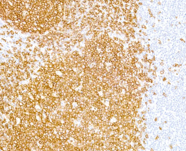
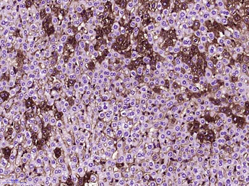
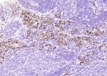
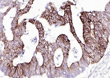
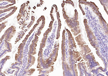
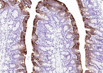
Detection of human CD20 by immunohistochemistry. Sample: FFPE section of human tonsil. Antibody: Purified mouse monoclonal anti-CD20 antibody used at a dilution of 1:500. Detection: DAB. Counterstain: IHC Hematoxylin (blue)
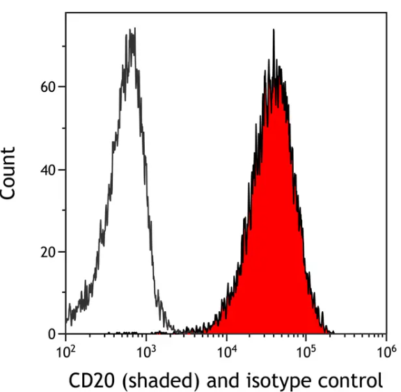
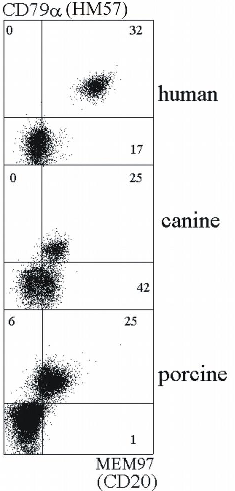
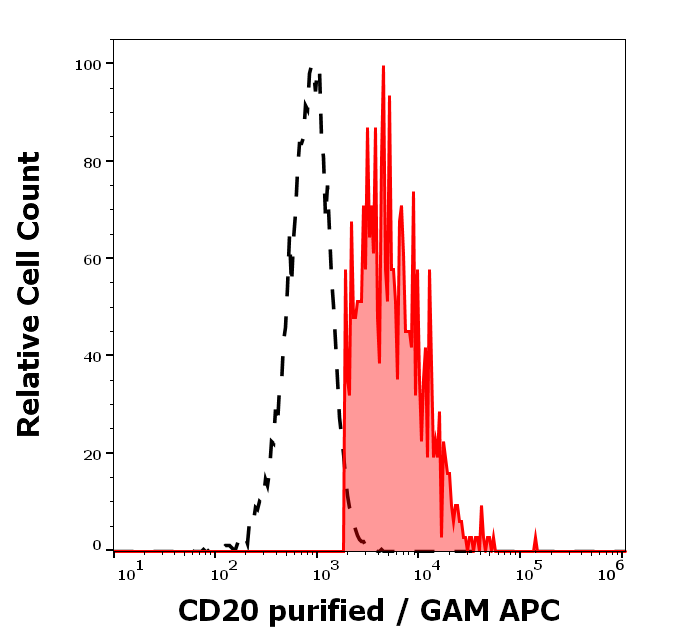
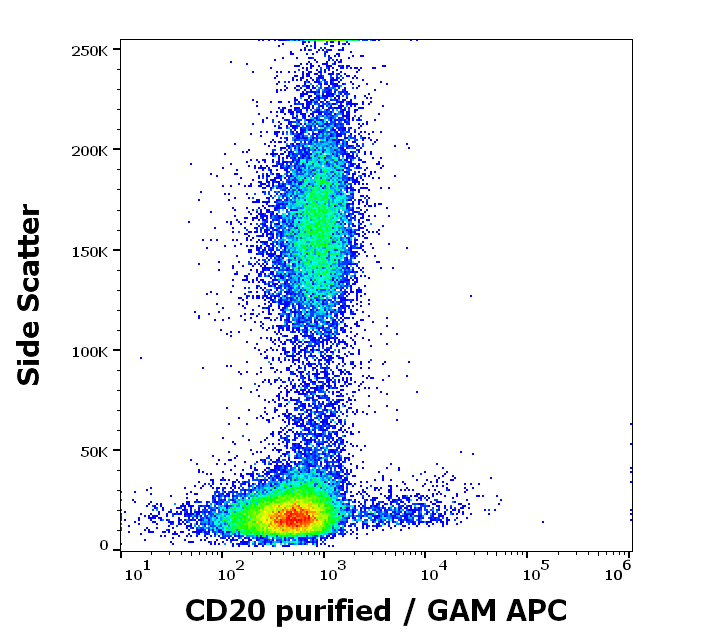
Detection of human CD20 (shaded) in Raji cells by flow cytometry. Antibody: Mouse anti-CD20 monoclonal antibody or isotype control (unshaded). Secondary: DyLight® 488-conjugated goat anti-mouse IgG
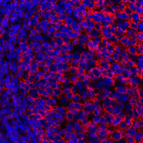
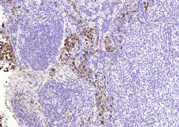
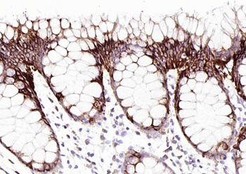
Detection of human CD20 by immunhistochemistry. Sample: FFPE section of human tonsil. Antibody: Mouse monoclonal anti-CD20 antibody used at 1:250. Secondary: DyLight® 594-conjugated goat anti-mouse IgG
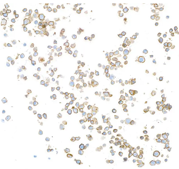
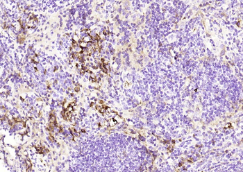
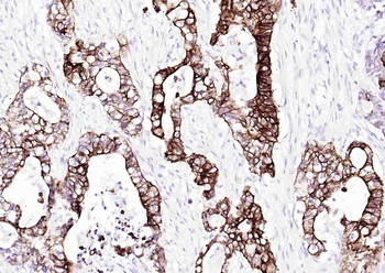
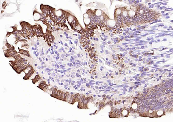
Detection of human CD20 by immunocytochemistry. Sample: FFPE section of VLN3G2 cells. Antibody: Purified mouse monoclonal anti-CD20 antibody used at a dilution of 1:500. Detection: DAB. Counterstain: IHC Hematoxylin (blue)
Quick Database Links
UniProt Details
− No UniProt data available
NCBI Reference Sequences
−Associated Accession Numbers
Curated reference sequences for the gene transcript and protein product| Protein | NP_068769.2 |
|---|
Documents Download
Datasheet
Product Information
Request a Document
Protocol Information
WB
Western Blot (IB, immunoblot)
IHC
Immunohistochemistry
FC
Flow Cytometry
IF
Immunofluorescence
CD20 Mouse Monoclonal Antibody (orb1520131)

- 0.0
Based on 0 reviews
Participating in our Biorbyt product reviews program enables you to support fellow scientists by sharing your firsthand experience with our products.
Login to Submit a ReviewAvailable Sizes
Select a size below
Choose Conjugation or Carrier Free Version
Free Secondary Antibody (20 ul)0/0
Please add an antibody product to your cart first.