You have no items in your shopping cart.
Featured
Description
Research Area
Cancer Biology
Images & Validation
−Item 1 of 2
| Tested Applications | FC, ICC, IF, IHC, IP, WB |
|---|---|
| Dilution Range | WB (1:1000), ICC/IF (1:100) |
| Reactivity | Human, Mouse, Rat |
| Application Notes |
Key Properties
−| Host | Mouse |
|---|---|
| Clonality | Monoclonal |
| Isotype | IgG2a |
| Clone No. | L51/82 (Formerly sold as S51-82) |
| Immunogen | Fusion protein amino acids 1-708 (full length) of rat MMP9 |
| Target | MMP9 |
| Molecular Weight | 82kDa |
| Purification | Protein G Purified |
| Conjugation | APC |
Storage & Handling
−| Storage | Conjugated antibodies should be stored according to the product label |
|---|---|
| Buffer/Preservatives | 95.46mM Phosphate, 2.48mM MES and 2mM EDTA |
| Concentration | 1 mg/ml |
| Expiration Date | 12 months from date of receipt. |
| Disclaimer | For research use only |
Alternative Names
−MMP9, MMP-9, Matrix metalloproteinase 9, CLG4B, 82kDa matrix metalloproteinase-9, Collagenease type 4 beta, GELB, Macrophage gelatinase, MANDP2, Type V collagenase, 92 kDa gelatinase, 92 kDa type IV collagenase, Gelatinase B
Similar Products
−
Lipocalin 2/NGAL Rabbit Polyclonal Antibody (APC) [orb995152]
IF
Bovine, Canine, Mouse, Porcine, Rabbit, Rat
Human
Rabbit
Polyclonal
APC
100 μlMMP9 Rabbit Polyclonal Antibody (APC) [orb1008318]
IF
Equine, Gallus, Mouse, Porcine, Rabbit, Rat, Sheep
Human
Rabbit
Polyclonal
APC
100 μl- Item 1 of 1
Adenomatous polyposis coli Antibody / APC [orb639712]
FACS, IF, WB
Human
Mouse
Monoclonal
Unconjugated
100 μg, 20 μg MMP9 Rabbit Polyclonal Antibody (APC-Cy7) [orb2416357]
IF
Equine, Gallus, Mouse, Porcine, Rabbit, Rat, Sheep
Human
Rabbit
Polyclonal
APC/Cy7
100 μl
Quality Guarantee
Explore bioreagents carefree to elevate your research. All our products are rigorously tested for performance. If a product does not perform as described on its datasheet, our scientific support team will provide expert troubleshooting, a prompt replacement, or a refund. For full details, please see our Terms & Conditions and Buying Guide. Contact us at [email protected].
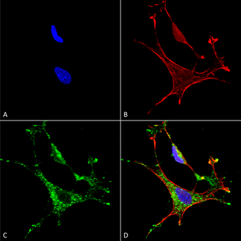
Immunocytochemistry/Immunofluorescence analysis using Mouse Anti-MMP9 Monoclonal Antibody, Clone L51/82. Tissue: Neuroblastoma cells (SH-SY5Y). Species: Human. Fixation: 4% PFA for 15 min. Primary Antibody: Mouse Anti-MMP9 Monoclonal Antibody at 1:50 for overnight at 4°C with slow rocking. Secondary Antibody: AlexaFluor 488 at 1:1000 for 1 hour at RT. Counterstain: Phalloidin-iFluor 647 (red) F-Actin stain; Hoechst (blue) nuclear stain at 1:800, 1.6mM for 20 min at RT. (A) Hoechst (blue) nuclear stain. (B) Phalloidin-iFluor 647 (red) F-Actin stain. (C) MMP9 Antibody (D) Composite.
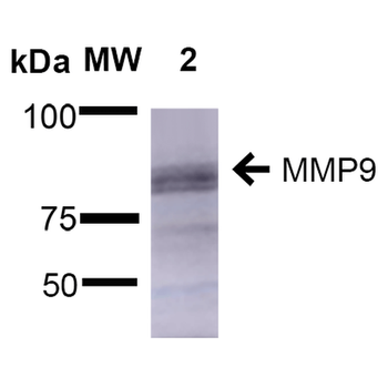
Western Blot analysis of Rat Brain showing detection of ~92 kDa and ~82 kDa (pro and active) MMP9 protein using Mouse Anti-MMP9 Monoclonal Antibody, Clone L51/82. Lane 1: Molecular Weight Ladder (MW). Lane 2: Rat Brain. Load: 15 μg. Block: 5% Skim Milk in 1X TBST. Primary Antibody: Mouse Anti-MMP9 Monoclonal Antibody at 1:1000 for 2 hours at RT. Secondary Antibody: Goat Anti-Mouse IgG: HRP at 1:2000 for 60 min at RT. Color Development: ECL solution for 5 min at RT. Predicted/Observed Size: ~92 kDa and ~82 kDa (pro and active).
Quick Database Links
UniProt Details
− No UniProt data available
NCBI Gene Details
− No NCBI Gene data available
NCBI Reference Sequences
−Associated Accession Numbers
Curated reference sequences for the gene transcript and protein product| Protein | NP_112317.1 |
|---|
Documents Download
Datasheet
Product Information
Request a Document
Protocol Information
WB
Western Blot (IB, immunoblot)
IHC
Immunohistochemistry
FC
Flow Cytometry
IF
Immunofluorescence
ICC
Immunocytochemistry
IP
Immunoprecipitation
MMP9 Antibody (APC) (orb149758)

- 0.0
Based on 0 reviews
Participating in our Biorbyt product reviews program enables you to support fellow scientists by sharing your firsthand experience with our products.
Login to Submit a ReviewAvailable Sizes
Select a size below
Choose Conjugation or Carrier Free Version
Free Secondary Antibody (20 ul)0/0
Please add an antibody product to your cart first.