You have no items in your shopping cart.
Description
Research Area
Cancer Biology, Immunology & Inflammation, Stem Cell & Developmental Biology
Images & Validation
−Item 1 of 4
| Tested Applications | FC, IHC-P, WB |
|---|---|
| Dilution Range | WB - 1:4000, IHC - 1:1000, IHC-P-Leica - 1:1000, FC - 1:25 |
| Reactivity | Human |
Key Properties
−| Antibody Type | Primary Antibody |
|---|---|
| Host | Mouse |
| Clonality | Monoclonal |
| Isotype | IgG1,k |
| Clone No. | B187EV1X3X3 |
| Immunogen | This MME antibody is generated from a mouse immunized with a KLH conjugated synthetic peptide between 472-505 amino acids from the Central region of human MME. |
| Target | MME {ECO:0000303|PubMed:27588448, ECO:0000312|HGNC:HGNC:7154} |
| Molecular Weight | 85514 Da |
| Conjugation | Unconjugated |
Storage & Handling
−| Storage | Maintain refrigerated at 2-8°C for up to 2 weeks. For long term storage store at -20°C in small aliquots to prevent freeze-thaw cycles |
|---|---|
| Form/Appearance | Purified monoclonal antibody supplied in PBS with 0.09% (W/V) sodium azide. This antibody is purified through a protein G column, followed by dialysis against PBS. |
| Expiration Date | 12 months from date of receipt. |
| Disclaimer | For research use only |
Alternative Names
−Neprilysin, 3.4.24.11, Atriopeptidase, Common acute lymphocytic leukemia antigen, CALLA, Enkephalinase, Neutral endopeptidase 24.11, NEP, Neutral endopeptidase, Skin fibroblast elastase, SFE, CD10, MME, EPN
Similar Products
−- Item 1 of 3
MME Antibody (Center) [orb1927088]
IHC-P, WB
Rabbit
Human, Mouse, Rat
Rabbit
Polyclonal
Unconjugated
50 μl, 100 μl - Item 1 of 2
- Item 1 of 2
- Item 1 of 4
MME Antibody (Center) [orb1788354]
IHC, WB
Human, Mouse, Rat
Rabbit
Polyclonal
Unconjugated
- Item 1 of 2
MMP12 Rabbit Polyclonal Antibody [orb214262]
IHC, WB
Human
Rabbit
Polyclonal
Unconjugated
30 μl, 100 μl, 200 μl, 50 μl


Quality Guarantee
Explore bioreagents carefree to elevate your research. All our products are rigorously tested for performance. If a product does not perform as described on its datasheet, our scientific support team will provide expert troubleshooting, a prompt replacement, or a refund. For full details, please see our Terms & Conditions and Buying Guide. Contact us at [email protected].
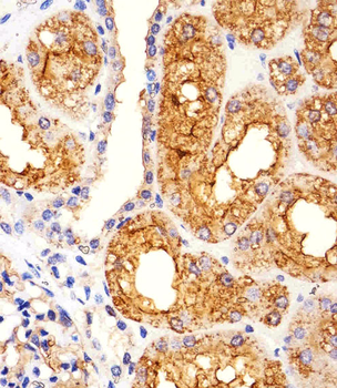
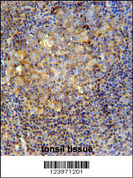
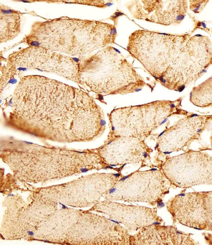
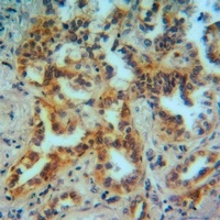
Immunohistochemical analysis of paraffin-embedded Human kidney section using Pink1. Diluted at 1:1000 dilution. A undiluted biotinylated goat polyvalent antibody was used as the secondary, followed by DAB staining.
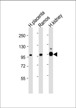
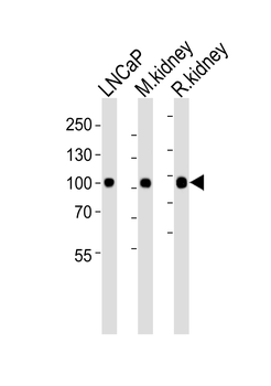
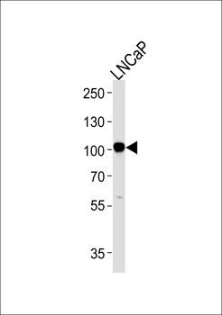
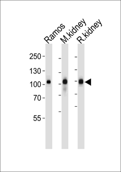
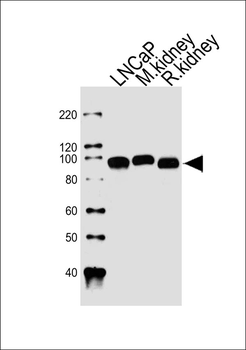
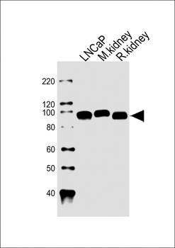
All lanes: Anti-MME Antibody (Center) at 1:4000 dilution. Lane 1: Human placenta lysate. Lane 2: Ramos whole cell lysate. Lane 3: Human kidney lysate. Lysates/proteins at 20 µg per lane. Secondary Goat Anti-mouse IgG, (H+L), Peroxidase conjugated at 1/10000 dilution. Predicted band size: 100 kDa. Blocking/Dilution buffer: 5% NFDM/TBST.
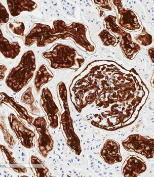
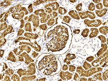
Immunohistochemical analysis of paraffin-embedded human kidney tissue was performed on the Leica BOND RXm. Tissue was fixed with formaldehyde at room temperature; antigen retrieval was by heat mediation with a EDTA buffer (pH9.0). Samples were incubated with primary antibody (1:1000) for 1 hours at room temperature. A undiluted biotinylated CRF Anti-Polyvalent HRP Polymer antibody was used as the secondary antibody.
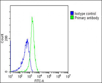
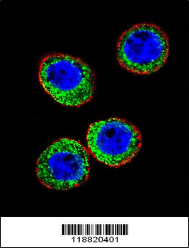
Overlay histogram showing Jurkat cells stained (green line). The cells were fixed with 2% paraformaldehyde (10 min). The cells were then icubated in 2% bovine serum albumin to block non-specific protein-protein interactions followed by the antibody (1:25 dilution) for 60 min at 37°C. The secondary antibody used was Goat-Anti-Mouse IgG, DyLight 488 Conjugated Highly Cross-Adsorbed (NH174309) at 1/200 dilution for 40 min at 37°C. Isotype control antibody (blue line) was mouse IgG1 (1 μg/1x10^6 cells) used under the same conditions. Acquisition of > 10000 events was performed.
Quick Database Links
Gene Symbol
MME {ECO:0000303|PubMed:27588448, ECO:0000312|HGNC:HGNC:7154}
UniProt
UniProt Details
− No UniProt data available
Documents Download
Datasheet
Product Information
Request a Document
Protocol Information
WB
Western Blot (IB, immunoblot)
IHC-P
Immunohistochemistry Paraffin
FC
Flow Cytometry
MME Antibody (Center) (orb1925550)

- 0.0
Based on 0 reviews
Participating in our Biorbyt product reviews program enables you to support fellow scientists by sharing your firsthand experience with our products.
Login to Submit a ReviewAvailable Sizes
Select a size below
Choose Conjugation or Carrier Free Version
Free Secondary Antibody (20 ul)0/0
Please add an antibody product to your cart first.