You have no items in your shopping cart.
Description
Images & Validation
−Item 1 of 3
| Tested Applications | IHC-P |
|---|---|
| Dilution Range | Immunohistochemistry (FFPE): 1-2ug/ml for 30 min at RT |
| Reactivity | Human |
| Application Notes |
Key Properties
−| Antibody Type | Primary Antibody |
|---|---|
| Host | Mouse |
| Clonality | Monoclonal |
| Isotype | Mouse IgG1, kappa |
| Clone No. | MTC719 |
| Immunogen | The Mitochondrial fraction of HeLa cells was used as the immunogen for this Mitochondrial antibody. |
| Purification | Protein G affinity chromatography |
| Conjugation | Unconjugated |
Storage & Handling
−| Storage | Maintain refrigerated at 2-8°C for up to 2 weeks. For long term storage store at -20°C in small aliquots to prevent freeze-thaw cycles. |
|---|---|
| Buffer/Preservatives | 0.2 mg/ml in 1X PBS with 0.1 mg/ml rAlbumin and 0.05% sodium azide |
| Expiration Date | 12 months from date of receipt. |
| Disclaimer | For research use only |
Similar Products
−- Item 1 of 19
COX4I1 Mouse Monoclonal Antibody (Mitochondrial Loading Control) [orb423160]
IF, IHC-Fr, IHC-P, WB
Mouse, Rat
Human, Mouse, Rat
Mouse
Monoclonal
Unconjugated
50 μl, 100 μl, 500 μl, 200 μg - Item 1 of 14
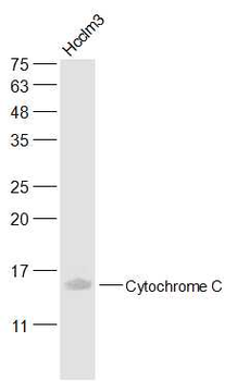
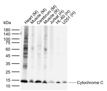
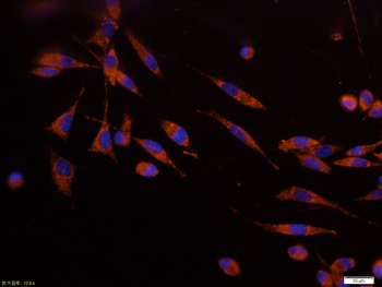
Cytochrome C Mouse Monoclonal Antibody [orb500962]
ICC, IF, IHC-Fr, IHC-P, WB
Mouse, Rat
Human, Mouse, Rat
Mouse
Monoclonal
Unconjugated
50 μl, 100 μl, 200 μl, 200 μg - Item 1 of 15
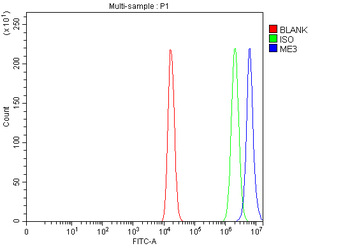
ME3 Rabbit Polyclonal Antibody [orb1972574]
ELISA, FC, IF, IHC, WB
Human, Monkey, Mouse, Rat
Rabbit
Polyclonal
Unconjugated
100 μg - Item 1 of 14
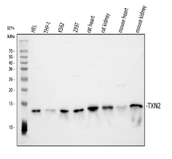
Thioredoxin 2/TXN2 Rabbit Polyclonal Antibody [orb570362]
ELISA, FC, ICC, IF, IHC, WB
Human, Mouse, Rat
Rabbit
Polyclonal
Unconjugated
100 μg - Item 1 of 14
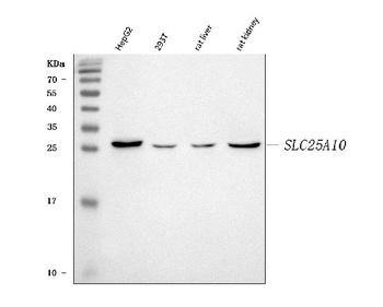
Mitochondrial dicarboxylate carrier/SLC25A10 Rabbit Polyclonal Antibody [orb1474851]
ELISA, IF, IHC, WB
Human, Mouse, Rat
Rabbit
Polyclonal
Unconjugated
100 μg

Quality Guarantee
Explore bioreagents carefree to elevate your research. All our products are rigorously tested for performance. If a product does not perform as described on its datasheet, our scientific support team will provide expert troubleshooting, a prompt replacement, or a refund. For full details, please see our Terms & Conditions and Buying Guide. Contact us at [email protected].
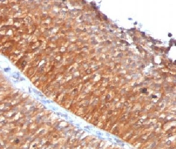
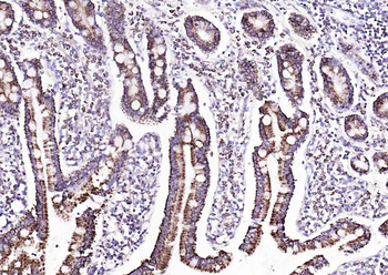
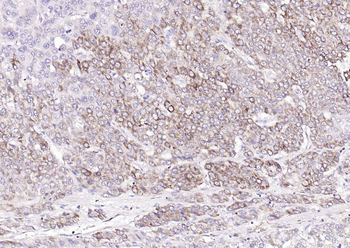
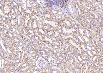
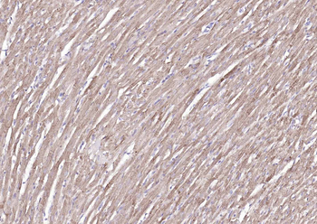
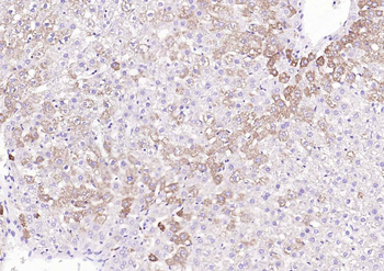
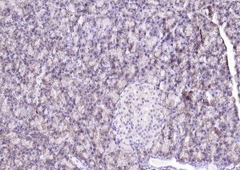
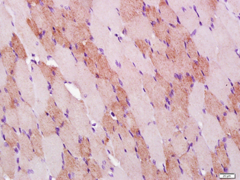
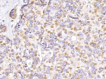
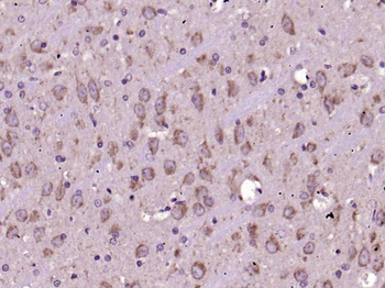
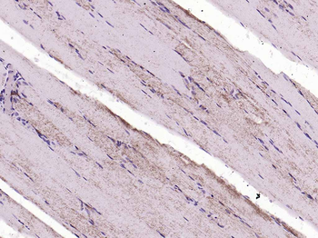
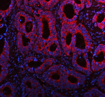
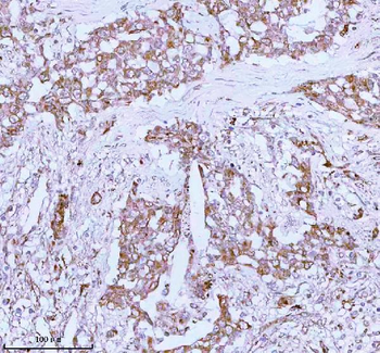
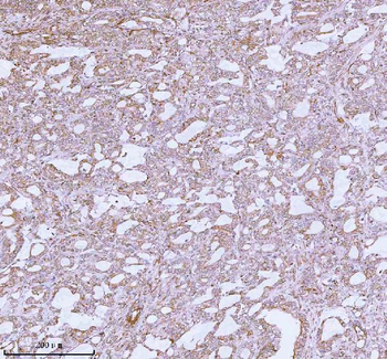
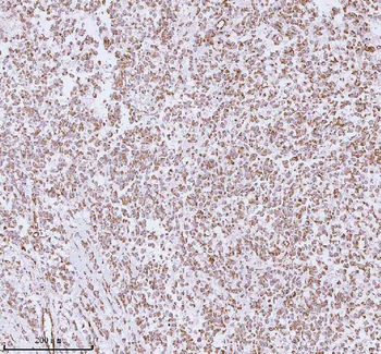
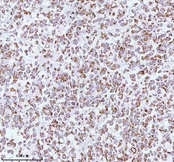
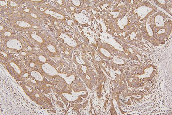
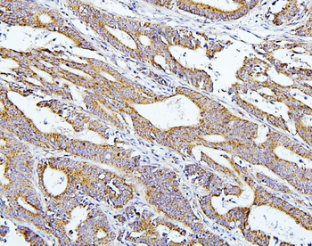
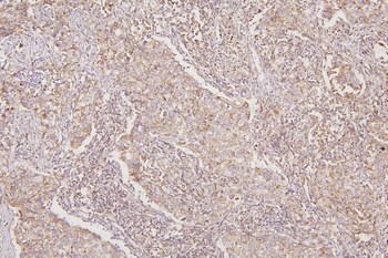
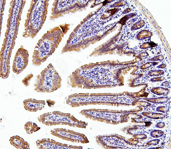
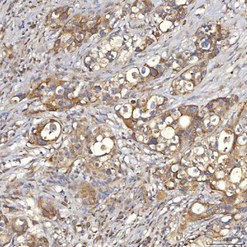
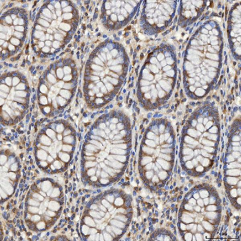
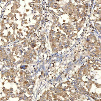
IHC testing of FFPE human bladder carcinoma with Mitochondrial antibody (clone MTC719).
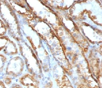
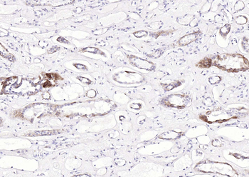
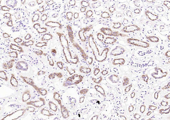
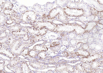
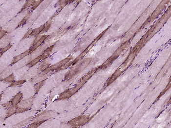
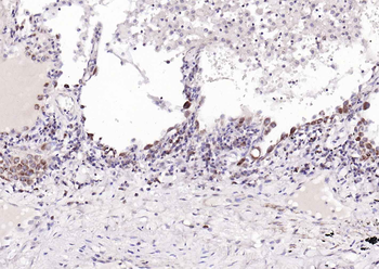
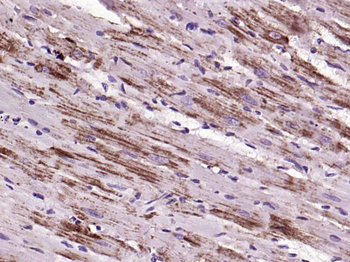
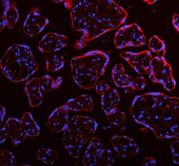
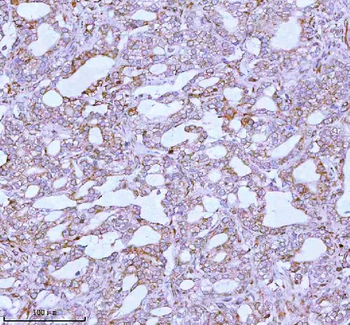
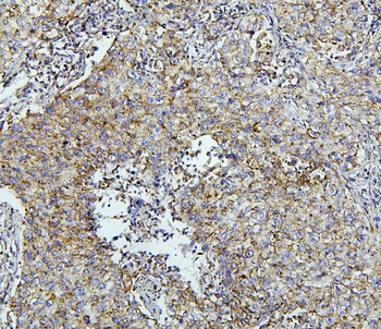
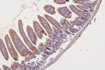
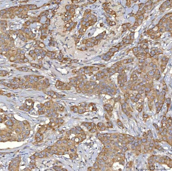
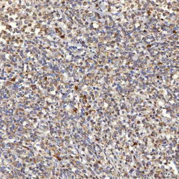
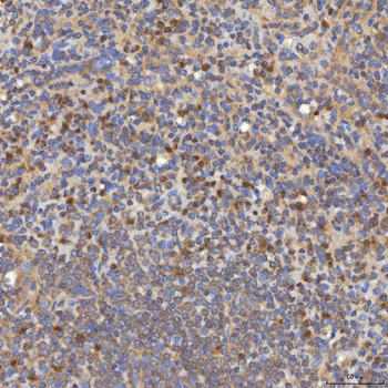
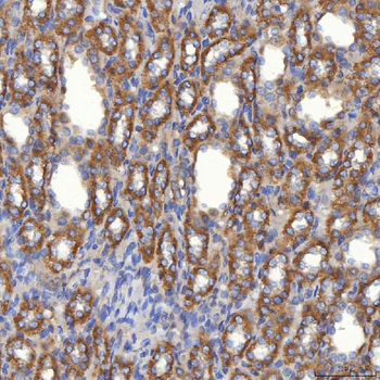
IHC testing of FFPE human renal cell carcinoma with Mitochondrial antibody (clone MTC719).
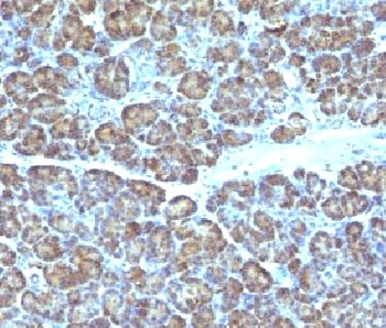
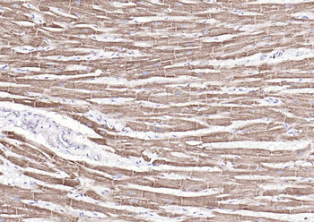
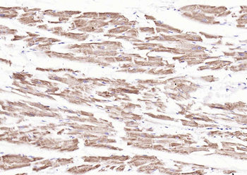
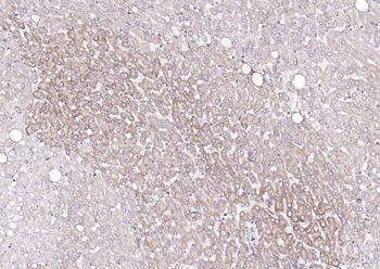
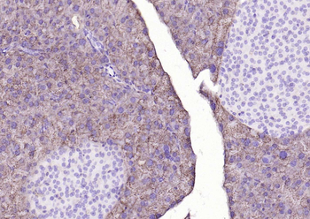
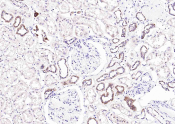
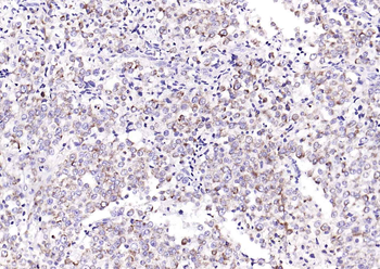
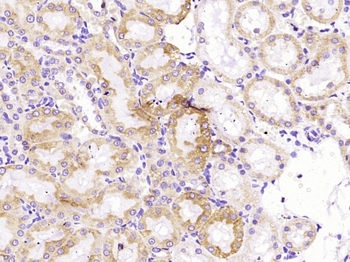
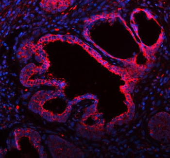
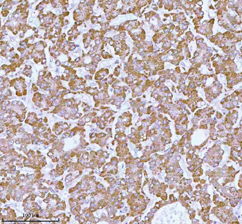
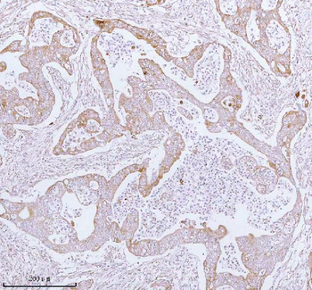
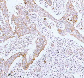
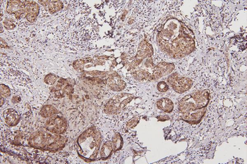
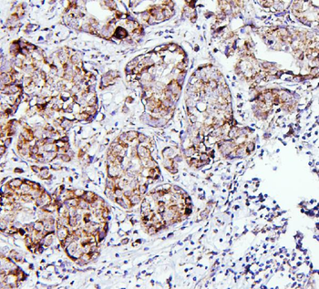
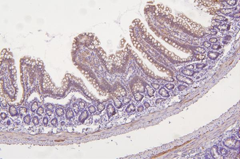
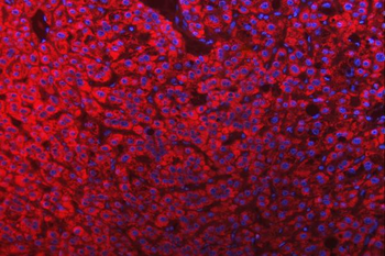
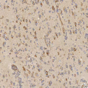
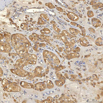
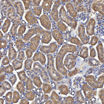
IHC testing of FFPE human pancreas with Mitochondrial antibody (clone MTC719).
Documents Download
Datasheet
Product Information
Request a Document
Mitochondrial Antibody (orb248672)

- 0.0
Based on 0 reviews
Participating in our Biorbyt product reviews program enables you to support fellow scientists by sharing your firsthand experience with our products.
Login to Submit a ReviewAvailable Sizes
Select a size below
Choose Conjugation or Carrier Free Version
Free Secondary Antibody (20 ul)0/0
Please add an antibody product to your cart first.