You have no items in your shopping cart.
Description
Research Area
Signal Transduction
Images & Validation
−Item 1 of 3
| Tested Applications | IHC-P, WB |
|---|---|
| Dilution Range | WB - 1:1000, IHC-P - 1:50-100 |
| Reactivity | Human |
Key Properties
−| Antibody Type | Primary Antibody |
|---|---|
| Host | Rabbit |
| Clonality | Polyclonal |
| Isotype | Rabbit IgG |
| Immunogen | This MEK3 (MAP2K3) antibody is generated from rabbits immunized with a KLH conjugated synthetic peptide between 15-45 amino acids from the N-terminal region of human MEK3 (MAP2K3). Antigen Region: 15-45 aa. |
| Target | MAP2K3 |
| Molecular Weight | 39318 Da |
| Conjugation | Unconjugated |
Storage & Handling
−| Storage | Maintain refrigerated at 2-8°C for up to 2 weeks. For long term storage store at -20°C in small aliquots to prevent freeze-thaw cycles |
|---|---|
| Form/Appearance | Purified polyclonal antibody supplied in PBS with 0.09% (W/V) sodium azide. This antibody is prepared by Saturated Ammonium Sulfate (SAS) precipitation followed by dialysis against PBS. |
| Expiration Date | 12 months from date of receipt. |
| Disclaimer | For research use only |
Alternative Names
−Dual specificity mitogen-activated protein kinase kinase 3, MAP kinase kinase 3, MAPKK 3, MAPK/ERK kinase 3, MEK 3, Stress-activated protein kinase kinase 2, SAPK kinase 2, SAPKK-2, SAPKK2, MAP2K3, MEK3, MKK3, PRKMK3, SKK2
Quality Guarantee
Explore bioreagents carefree to elevate your research. All our products are rigorously tested for performance. If a product does not perform as described on its datasheet, our scientific support team will provide expert troubleshooting, a prompt replacement, or a refund. For full details, please see our Terms & Conditions and Buying Guide. Contact us at [email protected].
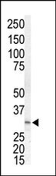
Western blot analysis of anti-MKK3 Pab in Jurkat cell lysate. MKK3 (arrow) was detected using purified Pab. Secondary HRP-anti-rabbit was used for signal visualization with chemiluminescence.
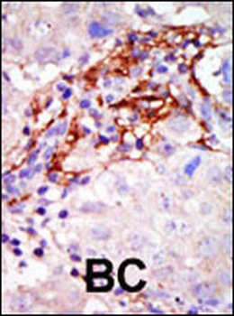
Formalin-fixed and paraffin-embedded human cancer tissue reacted with the primary antibody, which was peroxidase-conjugated to the secondary antibody, followed by AEC staining. This data demonstrates the use of this antibody for immunohistochemistry; clinical relevance has not been evaluated. BC = breast carcinoma; HC = hepatocarcinoma.
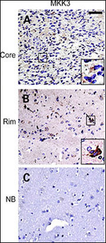
IHC of matched glioma core (top row) and rim (middle row) samples as well as normal brain control (bottom row) from a glioma invasion specific tissue microarray. Insets, individual tumor cells (black squares) are presented at higher magnification to facilitate interpretation of the staining pattern. Staining for MKK3 and pMKK3 exhibits strong signal in majority of invasive cells (B) compared with stationary cells from tumor core (A). Normal brain control shows only weak staining in reactive astrocytes (C).
Quick Database Links
UniProt Details
− No UniProt data available
NCBI Reference Sequences
−Associated Accession Numbers
Curated reference sequences for the gene transcript and protein product| Protein | NP_659731.1, NP_002747.2 |
|---|
Documents Download
Datasheet
Product Information
Request a Document
Protocol Information
WB
Western Blot (IB, immunoblot)
IHC-P
Immunohistochemistry Paraffin
MEK3 (MAP2K3) Antibody (N-term) (orb1928768)

- 0.0
Based on 0 reviews
Participating in our Biorbyt product reviews program enables you to support fellow scientists by sharing your firsthand experience with our products.
Login to Submit a ReviewAvailable Sizes
Select a size below
Choose Conjugation or Carrier Free Version
Free Secondary Antibody (20 ul)0/0
Please add an antibody product to your cart first.