You have no items in your shopping cart.
Description
Research Area
Metabolism Research
Images & Validation
−Item 1 of 4
| Tested Applications | DOT, FC, IHC-P, WB |
|---|---|
| Dilution Range | WB - 1:1000, IHC-P - 1:50-100, FC - 1:10-50, DB - 1:500 |
| Reactivity | Human |
Key Properties
−| Antibody Type | Primary Antibody |
|---|---|
| Host | Rabbit |
| Clonality | Polyclonal |
| Isotype | Rabbit IgG |
| Immunogen | This H4 antibody is generated from rabbits immunized with a KLH conjugated synthetic peptide between 1-30 amino acids from human H4. Antigen Region: 1-30 aa. |
| Target | H4C1 |
| Molecular Weight | 11367 Da |
| Conjugation | Unconjugated |
Storage & Handling
−| Storage | Maintain refrigerated at 2-8°C for up to 2 weeks. For long term storage store at -20°C in small aliquots to prevent freeze-thaw cycles |
|---|---|
| Form/Appearance | Purified polyclonal antibody supplied in PBS with 0.09% (W/V) sodium azide. This antibody is purified through a protein A column, followed by peptide affinity purification. |
| Expiration Date | 12 months from date of receipt. |
| Disclaimer | For research use only |
Alternative Names
−Histone H4, HIST1H4A, H4/A, H4FA
Similar Products
−Quality Guarantee
Explore bioreagents carefree to elevate your research. All our products are rigorously tested for performance. If a product does not perform as described on its datasheet, our scientific support team will provide expert troubleshooting, a prompt replacement, or a refund. For full details, please see our Terms & Conditions and Buying Guide. Contact us at [email protected].
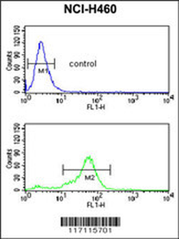
H4 Antibody (K20 [Me2]) flow cytometric analysis of NCI-H460 cells (bottom histogram) compared to a negative control cell (top histogram). FITC-conjugated goat-anti-rabbit secondary antibodies were used for the analysis.
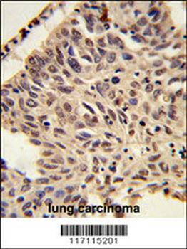
Formalin-fixed and paraffin-embedded human lung carcinoma reacted with PSMA4 Antibody (C-term), which was peroxidase-conjugated to the secondary antibody, followed by DAB staining. This data demonstrates the use of this antibody for immunohistochemistry; clinical relevance has not been evaluated.
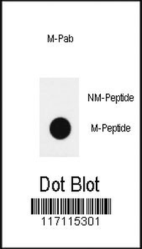
Dot blot analysis of anti-hH4-K20 (Methyl 2) methylation-specific Pab on nitrocellulose membrane. 50ng of methylation-peptide or Non methylation-peptide per dot were adsorbed. Antibody working concentrations are 0.5ug per ml.
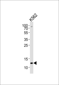
Western blot analysis of lysate from K562 cell line, using H4 Antibody (K20). Diluted at 1:1000 at each lane. A goat anti-rabbit IgG H&L (HRP) at 1:5000 dilution was used as the secondary antibody. Lysate at 35 ug per lane.
Quick Database Links
Gene Symbol
H4C1
UniProt
RefSeq (Protein):NP_003533.1, NP_003537.1, NP_003529.1, NP_003534.1, NP_003532.1, NP_003536.1, NP_003531.1, NP_003530.1, NP_003539.1, NP_068803.1, NP_778224.1, NP_003486.1, NP_001029249.1, NP_003535.1
UniProt Details
− No UniProt data available
NCBI Reference Sequences
−Associated Accession Numbers
Curated reference sequences for the gene transcript and protein productDocuments Download
Datasheet
Product Information
Request a Document
Protocol Information
WB
Western Blot (IB, immunoblot)
IHC-P
Immunohistochemistry Paraffin
FC
Flow Cytometry
DOT
Dot Blot
Me2-H4(K20) Antibody (orb1931072)

- 0.0
Based on 0 reviews
Participating in our Biorbyt product reviews program enables you to support fellow scientists by sharing your firsthand experience with our products.
Login to Submit a ReviewAvailable Sizes
Select a size below
Choose Conjugation or Carrier Free Version
Free Secondary Antibody (20 ul)0/0
Please add an antibody product to your cart first.