You have no items in your shopping cart.
Description
Research Area
Cell Biology
Images & Validation
−Item 1 of 4
| Tested Applications | FC, IF, IHC-P, WB |
|---|---|
| Dilution Range | IF - 1:10-50, WB - 1:2000, IHC-P - 1:10-50, FC - 1:25 |
| Reactivity | Human |
Key Properties
−| Host | Rabbit |
|---|---|
| Clonality | Polyclonal |
| Isotype | Rabbit IgG |
| Immunogen | This MDM2 antibody is generated from rabbits immunized with a KLH conjugated synthetic peptide between 141-176 amino acids from human MDM2. Antigen Region: 141-176 aa. |
| Target | MDM2 |
| Molecular Weight | 55233 Da |
| Conjugation | Unconjugated |
Storage & Handling
−| Storage | Maintain refrigerated at 2-8°C for up to 2 weeks. For long term storage store at -20°C in small aliquots to prevent freeze-thaw cycles |
|---|---|
| Form/Appearance | Purified polyclonal antibody supplied in PBS with 0.09% (W/V) sodium azide. This antibody is purified through a protein A column, followed by peptide affinity purification. |
| Expiration Date | 12 months from date of receipt. |
| Disclaimer | For research use only |
Alternative Names
−E3 ubiquitin-protein ligase Mdm2, 632-, Double minute 2 protein, Hdm2, Oncoprotein Mdm2, p53-binding protein Mdm2, MDM2
Similar Products
−- Item 1 of 3
Phospho-MDM2 (Ser166) Rabbit Polyclonal Antibody [orb6404]
FC, ICC, IF, IHC-Fr, IHC-P
Equine, Mouse, Rabbit, Rat
Human
Rabbit
Polyclonal
Unconjugated
50 μl, 100 μl, 200 μl - Item 1 of 3
MDM2 (Phospho-S166) Rabbit Polyclonal Antibody [orb216152]
IF, IHC, WB
Human, Mouse, Rat
Rabbit
Polyclonal
Unconjugated
30 μl, 100 μl, 200 μl, 50 μl - Item 1 of 2
MDM2 Antibody [orb675697]
ELISA, IF, IHC, WB
Human, Monkey, Mouse
Rabbit
Polyclonal
Unconjugated
100 μg, 50 μg - Item 1 of 1
Phospho-MDM2 (Ser166) Recombinant Rabbit Monoclonal Antibody [orb2561450]
ICC, IF, IHC-Fr, IHC-P, WB
Human
Human
Rabbit
Recombinant
Unconjugated
50 μl, 100 μl, 25 μl 
Phospho-MDM2 (Ser166) Rabbit Polyclonal Antibody (FITC) [orb9252]
FC, ICC, IF
Equine, Mouse, Rabbit, Rat
Human
Rabbit
Polyclonal
FITC
100 μl


Quality Guarantee
Explore bioreagents carefree to elevate your research. All our products are rigorously tested for performance. If a product does not perform as described on its datasheet, our scientific support team will provide expert troubleshooting, a prompt replacement, or a refund. For full details, please see our Terms & Conditions and Buying Guide. Contact us at [email protected].
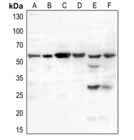
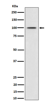
Anti-MDM2 Antibody (S166) at1:2000 dilution + CCRF-CEM whole cell lysate.Lysates/proteins at 20 µg per lane. Secondary Goat Anti-Rabbit IgG, (H+L), Peroxidase conjugated at 1/10000 dilution. Predicted band size: 55 kDa. Blocking/Dilution buffer: 5% NFDM/TBST.
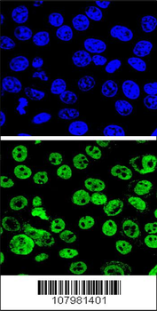
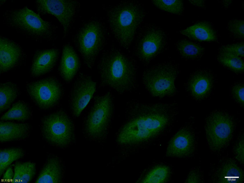
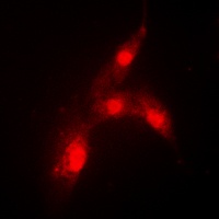
Confocal immunofluorescent analysis of MDM2 Antibody (S166) with Hela cell followed by Alexa Fluor 488-conjugated goat anti-rabbit lgG (green). DAPI was used to stain the cell nuclear (blue).
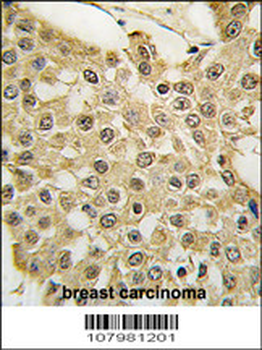
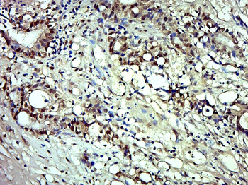
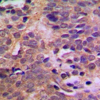
Formalin-fixed and paraffin-embedded human breast carcinoma tissue reacted with the MDM2 Antibody (S166), which was peroxidase-conjugated to the secondary antibody, followed by DAB staining. This data demonstrates the use of this antibody for immunohistochemistry; clinical relevance has not been evaluated.
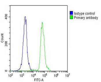
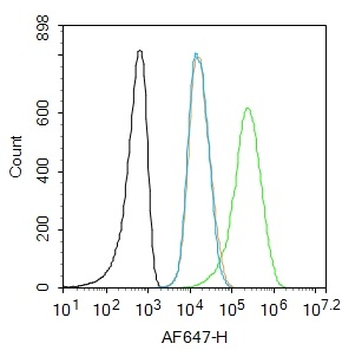
Overlay histogram showing THP-1 cells (green line). The cells were fixed with 2% paraformaldehyde (10 min) and then permeabilized with 90% methanol for 10 min. The cells were then icubated in 2% bovine serum albumin to block non-specific protein-protein interactions followed by the antibody (1:25 dilution) for 60 min at 37°C. The secondary antibody used was Goat-Anti-Rabbit IgG, DyLight 488 Conjugated Highly Cross-Adsorbed at 1/200 dilution for 40 min at 37°C. Isotype control antibody (blue line) was rabbit IgG (1 μg/1x10^6 cells) used under the same conditions. Acquisition of > 10000 events was performed.
Quick Database Links
UniProt Details
− No UniProt data available
NCBI Reference Sequences
−Associated Accession Numbers
Curated reference sequences for the gene transcript and protein product| Protein | NP_002383.2, NP_001265391.1, NP_001138811.1 |
|---|
Documents Download
Datasheet
Product Information
Request a Document
Protocol Information
WB
Western Blot (IB, immunoblot)
IHC-P
Immunohistochemistry Paraffin
FC
Flow Cytometry
IF
Immunofluorescence
MDM2 Antibody (S166) (orb1937385)

- 0.0
Based on 0 reviews
Participating in our Biorbyt product reviews program enables you to support fellow scientists by sharing your firsthand experience with our products.
Login to Submit a ReviewAvailable Sizes
Select a size below
Free Secondary Antibody (20 ul)0/0
Please add an antibody product to your cart first.