You have no items in your shopping cart.
Description
Research Area
Immunology & Inflammation
Images & Validation
−Item 1 of 6
| Tested Applications | ELISA, WB |
|---|---|
| Reactivity | Human |
Key Properties
−| Antibody Type | Primary Antibody |
|---|---|
| Host | Rabbit |
| Clonality | Polyclonal |
| Immunogen | Produced from sera of rabbits pre-immunized with highly pure (>98%) recombinant hMCP 1/MCAF (human Macrophage/Monocyte chemotactic protein-1). |
| Target | CCL2 |
| Purification | Anti-hMCP-1/MCAF specific antibody was purified by affinity chromatography employing immobilized hMCP-1/MCAF matrix. |
| Conjugation | Unconjugated |
Storage & Handling
−| Storage | Maintain refrigerated at 2-8°C for up to 2 weeks. For long term storage store at -20°C in small aliquots to prevent freeze-thaw cycles. |
|---|---|
| Form/Appearance | Lyophilized |
| Concentration | batch dependent |
| Expiration Date | 12 months from date of receipt. |
| Disclaimer | For research use only |
Alternative Names
−HC11, MCAF, MCP1, MCP-1, SCYA2, GDCF-2, SMC-CF, HSMCR30, C-C motif chemokine 2, HC11
Similar Products
−- Item 1 of 8
- Item 1 of 7
MCP1 Rabbit Polyclonal Antibody [orb323291]
ELISA, ICC, IF, IHC-P, WB
Human, Mouse, Porcine, Rat
Rabbit
Polyclonal
Unconjugated
100 μg - Item 1 of 7
CCL2 Antibody [orb97456]
FC, ICC, IHC, WB
Human, Monkey, Mouse, Rat
Mouse
Monoclonal
Unconjugated
100 μl - Item 1 of 1
MCP1 Rabbit Polyclonal Antibody [orb13563]
ELISA, WB
Human
Mouse, Rat
Rabbit
Polyclonal
Unconjugated
50 μl, 100 μl, 200 μl - Item 1 of 1
Human Monocyte Chemotactic Protein 1 (MCP1) ELISA Kit [orb779110]
Human
15.63-1000 pg/mL
6.4 pg/mL
48 T, 96 T
Quality Guarantee
Explore bioreagents carefree to elevate your research. All our products are rigorously tested for performance. If a product does not perform as described on its datasheet, our scientific support team will provide expert troubleshooting, a prompt replacement, or a refund. For full details, please see our Terms & Conditions and Buying Guide. Contact us at [email protected].
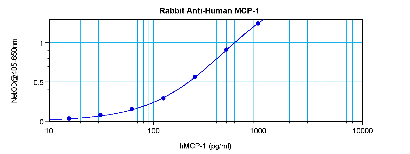
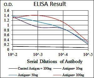
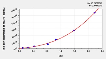
To detect hMCP-1 (MCAF) by sandwich ELISA (using 100 ul/well antibody solution) a concentration of 0.5 - 2.0 ug/ml of this antibody is required. This antigen affinity purified antibody, in conjunction with Anti-Human MCP-1 (MCAF) (orb1272403) as a detection antibody, allows the detection of at least 0.2 - 0.4 ng/well of recombinant hMCP-1 (MCAF).
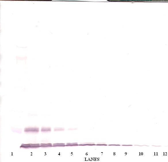
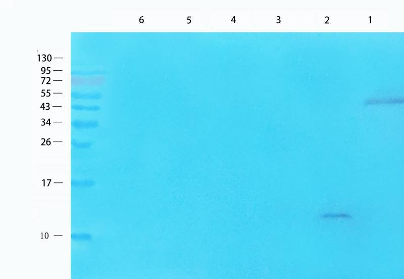
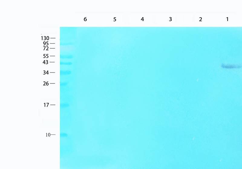
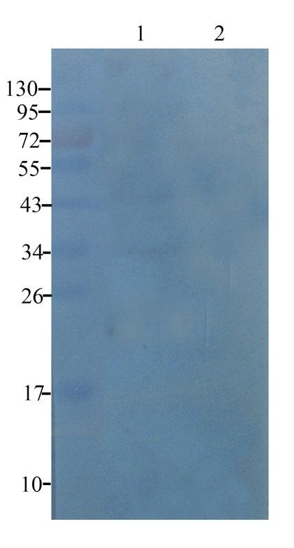
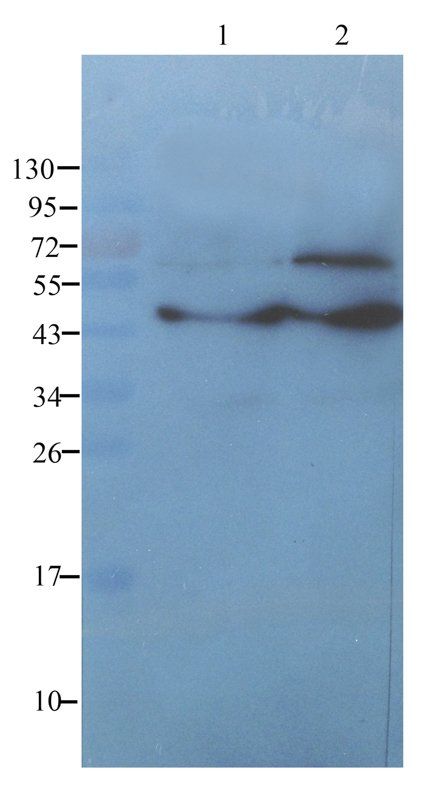
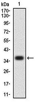
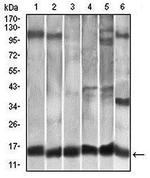
To detect hMCP-1 (MCAF) by Western Blot analysis this antibody can be used at a concentration of 0.1-0.2 ug/ml. Used in conjunction with compatible secondary reagents the detection limit for recombinant hMCP-1 (MCAF) is 1.5-3.0 ng/lane, under either reducing or non-reducing conditions.
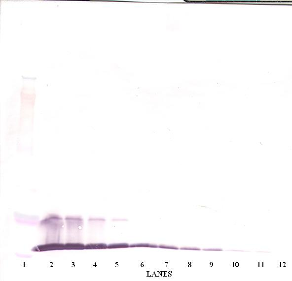
To detect hMCP-1 (MCAF) by Western Blot analysis this antibody can be used at a concentration of 0.1-0.2 ug/ml. Used in conjunction with compatible secondary reagents the detection limit for recombinant hMCP-1 (MCAF) is 1.5-3.0 ng/lane, under either reducing or non-reducing conditions.
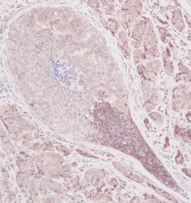
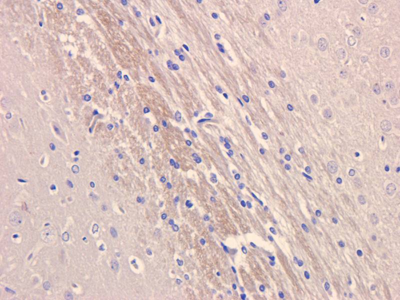
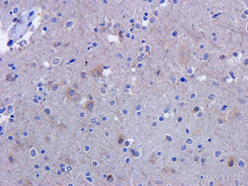
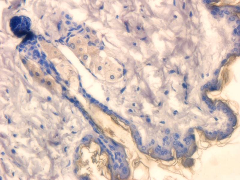
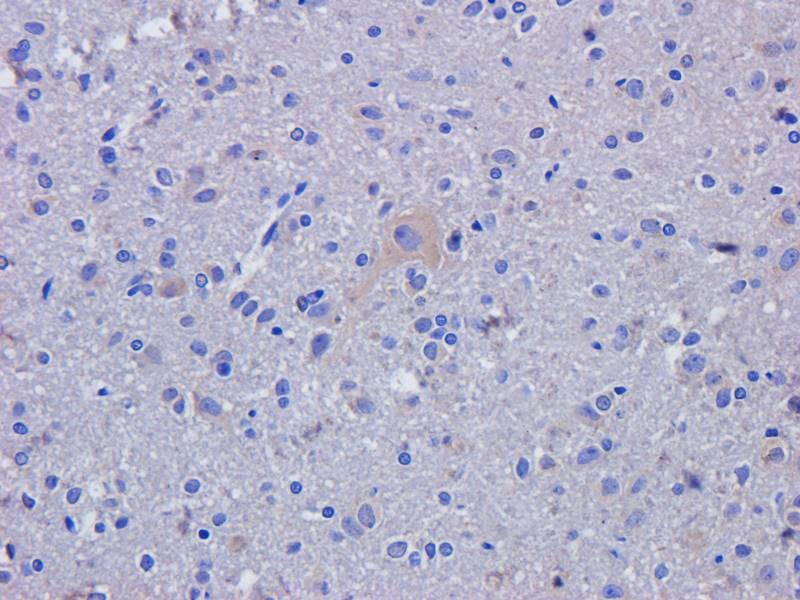
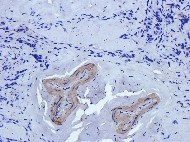
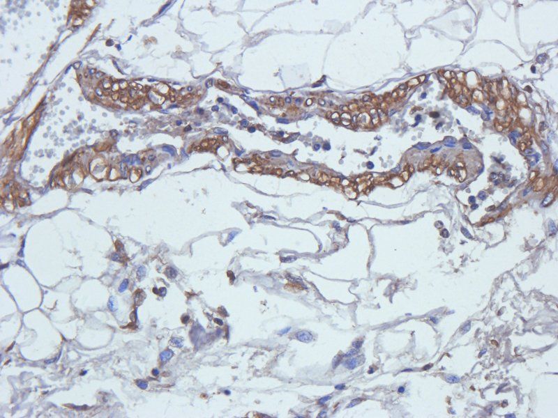
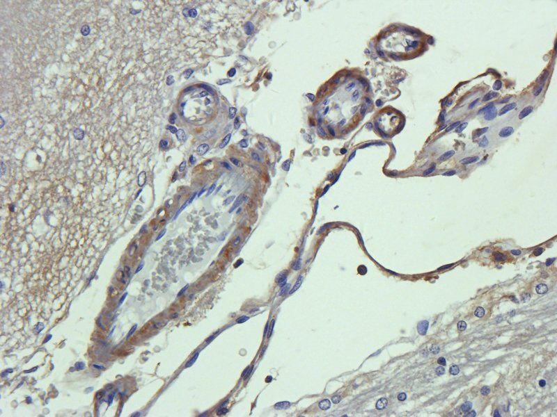
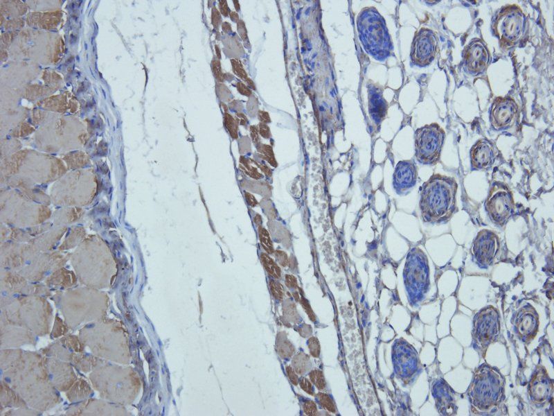
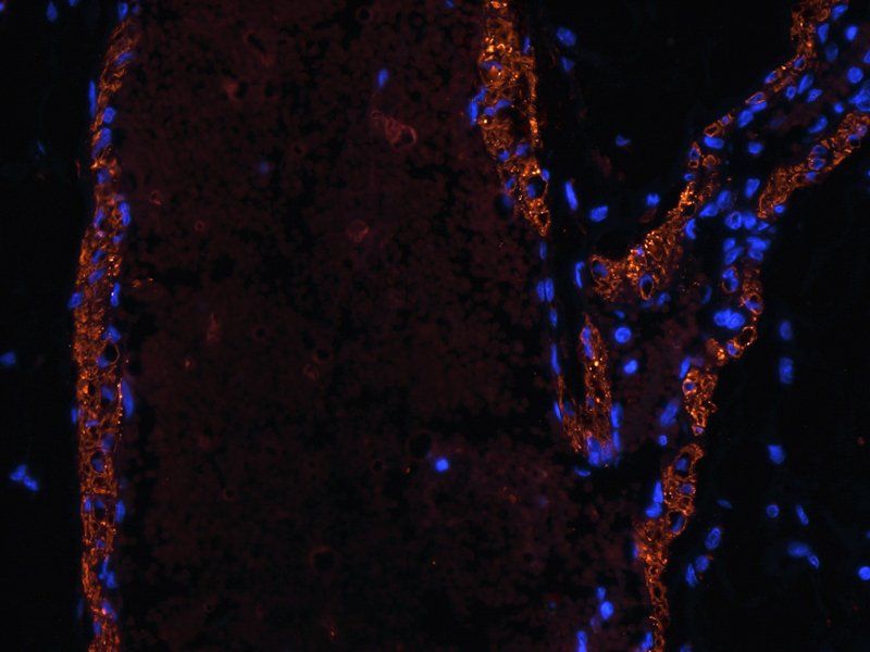
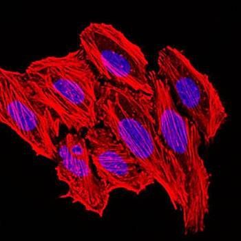
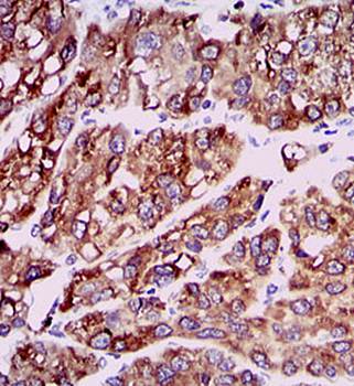
This antibody stained formalin-fixed, paraffin-embedded sections of human breast invasive ductal carcinoma. The recommended concentration is 2.5 ug/ml and a two-hour incubation at room temperature. An HRP-labeled polymer detection system was used with a DAB chromogen. Heat induced antigen retrieval with a pH 6.0 Sodium Citrate buffer is recommended. Optimal concentrations and conditions may vary.
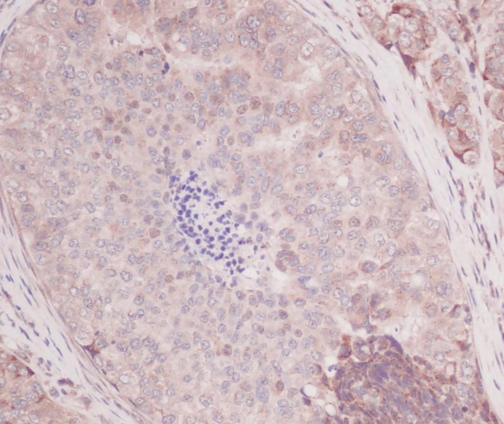
This antibody stained formalin-fixed, paraffin-embedded sections of human breast invasive ductal carcinoma. The recommended concentration is 2.5 ug/ml and a two-hour incubation at room temperature. An HRP-labeled polymer detection system was used with a DAB chromogen. Heat induced antigen retrieval with a pH 6.0 Sodium Citrate buffer is recommended. Optimal concentrations and conditions may vary.
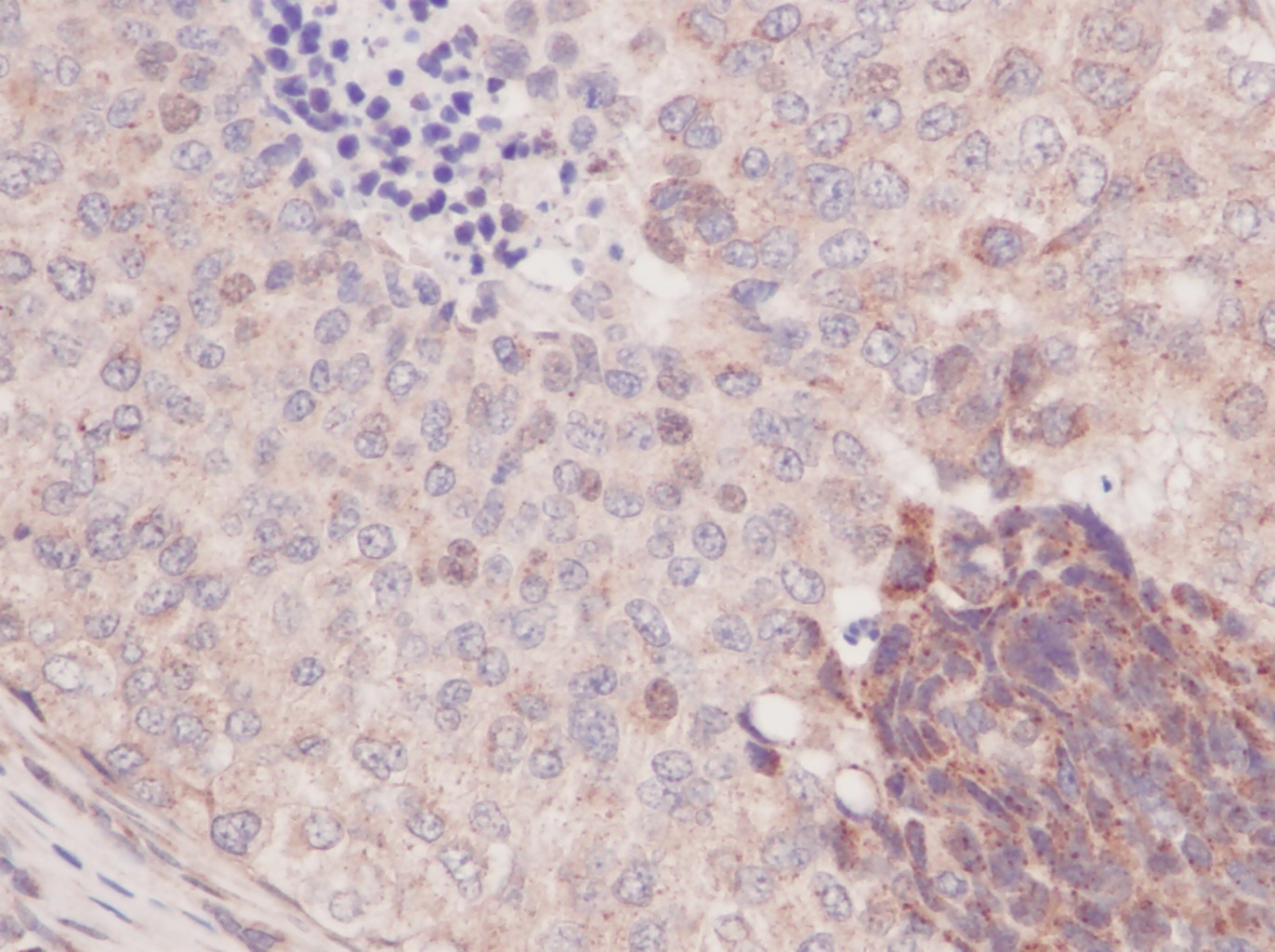
This antibody stained formalin-fixed, paraffin-embedded sections of human breast invasive ductal carcinoma. The recommended concentration is 2.5 ug/ml and a two-hour incubation at room temperature. An HRP-labeled polymer detection system was used with a DAB chromogen. Heat induced antigen retrieval with a pH 6.0 Sodium Citrate buffer is recommended. Optimal concentrations and conditions may vary.
Documents Download
Datasheet
Product Information
Request a Document
Protocol Information
WB
Western Blot (IB, immunoblot)
ELISA
Enzyme-linked Immunosorbent Assay (EIA)
CCL2 Antibody (orb1272404)

- 0.0
Based on 0 reviews
Participating in our Biorbyt product reviews program enables you to support fellow scientists by sharing your firsthand experience with our products.
Login to Submit a ReviewAvailable Sizes
Select a size below
Free Secondary Antibody (20 ul)0/0
Please add an antibody product to your cart first.