You have no items in your shopping cart.
Featured
Description
Research Area
Cell Biology
Images & Validation
−Item 1 of 2
| Tested Applications | ICC, IF, IP, WB |
|---|---|
| Dilution Range | WB (1:1000), ICC/IF (1:500) |
| Reactivity | Human, Mouse, Rabbit |
| Application Notes |
Key Properties
−| Host | Rat |
|---|---|
| Clonality | Monoclonal |
| Isotype | IgG1 |
| Clone No. | GL2A7 |
| Immunogen | Purified preparation of mouse liver lysosomal membranes |
| Target | LAMP2 |
| Molecular Weight | 100-110kDa |
| Purification | Protein G Purified |
| Conjugation | Biotin |
Storage & Handling
−| Storage | Conjugated antibodies should be stored according to the product label |
|---|---|
| Buffer/Preservatives | 136.36mM Ethanolamine, 133.23 mM Chlorides, 9.55mM Phosphates, 9.55mM Sodium Bicarbonate |
| Concentration | 1 mg/ml |
| Expiration Date | 12 months from date of receipt. |
| Disclaimer | For research use only |
Alternative Names
−LAMP2, LAMP-2, Lamp2C, Lysosome-associated membrane glycoprotein 2, Lysosome-associated membrane protein 2, CD107b, CD107 antigen-like family member B, Lysosomal membrane glycoprotein type B, LGP-B, Igp110, Igp2, LampB, MAC3
Similar Products
−- Item 1 of 1
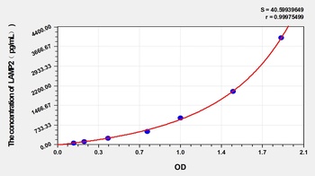
Rabbit Lysosomal Associated Membrane Protein 2 (LAMP2) ELISA Kit [orb1173550]
Rabbit
62.5-4000 pg/mL
23.7 pg/mL
48 T, 96 T - Item 1 of 2
LAMP1 Antibody (Biotin) [orb147228]
ICC, IF, IP, WB
Hamster, Human, Mouse, Rat
Mouse
Monoclonal
Biotin
100 μg 
LAMP2 Rabbit Polyclonal Antibody (Biotin) [orb458470]
FC, ICC, IF, IHC-Fr, IHC-P, IP, WB
Bovine, Canine, Equine, Porcine, Rabbit
Human, Mouse, Rat
Rabbit
Polyclonal
Biotin
100 μl
Quality Guarantee
Explore bioreagents carefree to elevate your research. All our products are rigorously tested for performance. If a product does not perform as described on its datasheet, our scientific support team will provide expert troubleshooting, a prompt replacement, or a refund. For full details, please see our Terms & Conditions and Buying Guide. Contact us at [email protected].
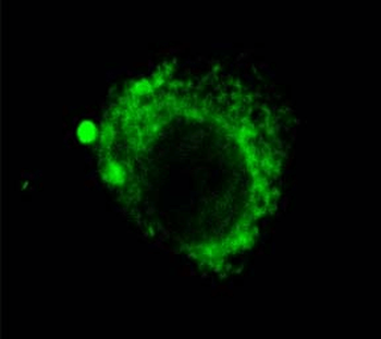
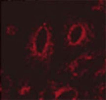
Immunocytochemistry/Immunofluorescence analysis using Rat Anti-LAMP2 Monoclonal Antibody, Clone GL2A7. Tissue: Corneal Endothelial Cell (CEC). Species: Rabbit. Primary Antibody: Rat Anti-LAMP2 Monoclonal Antibody at 1:1000. Secondary Antibody: FITC Goat Anti-Rat (green).
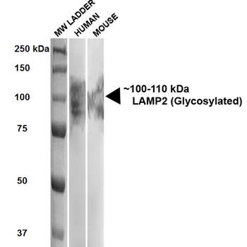
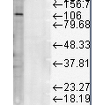
Western Blot analysis of Human, Mouse HEK293 and 3T3NIH cell lysates showing detection of ~100-110 kDa LAMP2 protein using Rat Anti-LAMP2 Monoclonal Antibody, Clone GL2A7. Lane 1: MW ladder. Lane 2: Human HEK293 lysate (20 μg). Lane 3: Mouse 3T3NIH lysate (10 μg). Block: 5% milk + TBST for 1 hour at RT. Primary Antibody: Rat Anti-LAMP2 Monoclonal Antibody at 1:500 for 1 hour at RT. Secondary Antibody: HRP Goat Anti-Rat at 1:100 for 1 hour at RT. Color Development: TMB solution for 5 min at RT. Predicted/Observed Size: ~100-110 kDa.
Quick Database Links
UniProt Details
− No UniProt data available
NCBI Gene Details
− No NCBI Gene data available
NCBI Reference Sequences
−Associated Accession Numbers
Curated reference sequences for the gene transcript and protein product| Protein | NP_001017959.1 |
|---|
Documents Download
Datasheet
Product Information
Request a Document
Protocol Information
WB
Western Blot (IB, immunoblot)
IF
Immunofluorescence
ICC
Immunocytochemistry
IP
Immunoprecipitation
LAMP2 Antibody (Biotin) (orb147245)

- 0.0
Based on 0 reviews
Participating in our Biorbyt product reviews program enables you to support fellow scientists by sharing your firsthand experience with our products.
Login to Submit a ReviewAvailable Sizes
Select a size below
Choose Conjugation or Carrier Free Version
Free Secondary Antibody (20 ul)0/0
Please add an antibody product to your cart first.