You have no items in your shopping cart.
Featured
Description
Research Area
Cancer Biology, Neuroscience, Pharmacology & Drug Discovery
Images & Validation
−Item 1 of 4
| Tested Applications | AM, ICC, IF, IHC, WB |
|---|---|
| Dilution Range | WB (1:1000), IHC (1:1000), ICC/IF (1:100) |
| Reactivity | Human, Mouse, Rat |
| Application Notes |
Key Properties
−| Host | Mouse |
|---|---|
| Clonality | Monoclonal |
| Isotype | IgG1 |
| Clone No. | N43/6 (Formerly sold as S43-6) |
| Immunogen | Fusion protein amino acids 2-77 of human KCNQ4 |
| Target | KCNQ4 |
| Molecular Weight | 77kDa |
| Purification | Protein G Purified |
| Conjugation | PerCP |
Storage & Handling
−| Storage | Conjugated antibodies should be stored according to the product label |
|---|---|
| Buffer/Preservatives | 95.46mM Phosphate, 2.48mM MES and 2mM EDTA |
| Concentration | 1 mg/ml |
| Expiration Date | 12 months from date of receipt. |
| Disclaimer | For research use only |
Alternative Names
−KCNQ4, Kv7.4, KVLQT4, Potassium voltage-gated channel subfamily KQT member 4, Voltage-gated potassium channel subunit Kv7.4, KQT-like 4, Potassium channel subunit alpha KvLQT4, Potassium voltage-gated channel KQT-like subfamily member 4, DFNA2, KCNQ4_HUMAN, Kcnq4, KCNQ 4, Potassium channel KQT like 4, Potassium voltage gated channel KQT like protein 4, Voltage gated potassium channel subunit Kv7.4
Quality Guarantee
Explore bioreagents carefree to elevate your research. All our products are rigorously tested for performance. If a product does not perform as described on its datasheet, our scientific support team will provide expert troubleshooting, a prompt replacement, or a refund. For full details, please see our Terms & Conditions and Buying Guide. Contact us at [email protected].
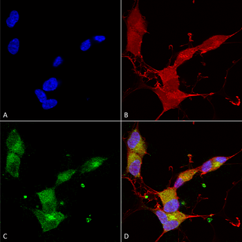
Immunocytochemistry/Immunofluorescence analysis using Mouse Anti-KCNQ4 Monoclonal Antibody, Clone N43/6. Tissue: Neuroblastoma cells (SH-SY5Y). Species: Human. Fixation: 4% PFA for 15 min. Primary Antibody: Mouse Anti-KCNQ4 Monoclonal Antibody at 1:100 for overnight at 4°C with slow rocking. Secondary Antibody: AlexaFluor 488 at 1:1000 for 1 hour at RT. Counterstain: Phalloidin-iFluor 647 (red) F-Actin stain; Hoechst (blue) nuclear stain at 1:800, 1.6mM for 20 min at RT. (A) Hoechst (blue) nuclear stain. (B) Phalloidin-iFluor 647 (red) F-Actin stain. (C) KCNQ4 Antibody (D) Composite.
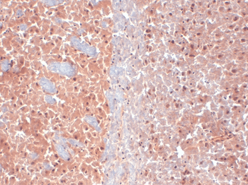
Immunohistochemistry analysis using Mouse Anti-KCNQ4 Monoclonal Antibody, Clone N43/6. Tissue: frozen brain section. Species: Mouse. Fixation: 10% Formalin Solution for 12-24 hours at RT. Primary Antibody: Mouse Anti-KCNQ4 Monoclonal Antibody at 1:1000 for 1 hour at RT. Secondary Antibody: HRP/DAB Detection System: Biotinylated Goat Anti-Mouse, Streptavidin Peroxidase, DAB Chromogen (brown) for 30 minutes at RT. Counterstain: Mayer Hematoxylin (purple/blue) nuclear stain at 250-500 μl for 5 minutes at RT.
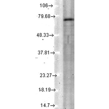
Western Blot analysis of Rat tissue lysate showing detection of KCNQ4 protein using Mouse Anti-KCNQ4 Monoclonal Antibody, Clone N43/6. Load: 15 μg. Block: 1.5% BSA for 30 minutes at RT. Primary Antibody: Mouse Anti-KCNQ4 Monoclonal Antibody at 1:1000 for 2 hours at RT. Secondary Antibody: Sheep Anti-Mouse IgG: HRP for 1 hour at RT.
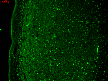
Immunohistochemistry analysis using Mouse Anti-KCNQ4 Monoclonal Antibody, Clone N43/6. Tissue: hippocampus. Species: Human. Fixation: Bouin's Fixative and paraffin-embedded. Primary Antibody: Mouse Anti-KCNQ4 Monoclonal Antibody at 1:1000 for 1 hour at RT. Secondary Antibody: FITC Goat Anti-Mouse (green) at 1:50 for 1 hour at RT.
Quick Database Links
UniProt Details
− No UniProt data available
NCBI Gene Details
− No NCBI Gene data available
NCBI Reference Sequences
−Associated Accession Numbers
Curated reference sequences for the gene transcript and protein product| Protein | NP_004691.2 |
|---|
Documents Download
Datasheet
Product Information
Request a Document
Protocol Information
WB
Western Blot (IB, immunoblot)
IHC
Immunohistochemistry
IF
Immunofluorescence
ICC
Immunocytochemistry
KCNQ4 Antibody (PerCP) (orb148318)

- 0.0
Based on 0 reviews
Participating in our Biorbyt product reviews program enables you to support fellow scientists by sharing your firsthand experience with our products.
Login to Submit a ReviewAvailable Sizes
Select a size below
Choose Conjugation or Carrier Free Version
Free Secondary Antibody (20 ul)0/0
Please add an antibody product to your cart first.