You have no items in your shopping cart.
Featured
Description
Research Area
Cell Biology
Images & Validation
−
Item 1 of 5
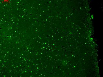
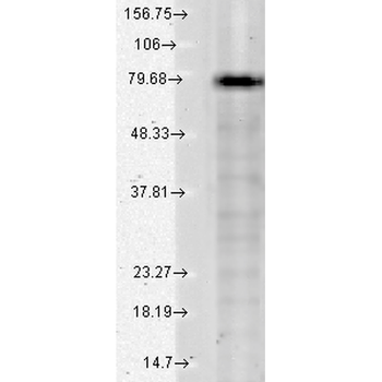
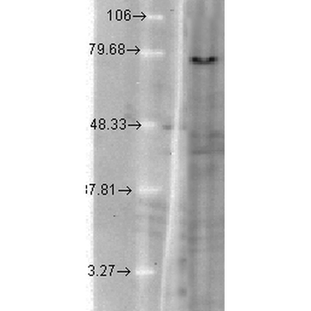
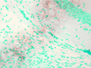
| Tested Applications | AM, ICC, IF, IHC, IP, WB |
|---|---|
| Dilution Range | WB (1:1000), IHC (1:1000), ICC/IF (1:100) |
| Reactivity | Hamster, Human, Mouse, Rat |
| Application Notes |
Key Properties
−| Host | Mouse |
|---|---|
| Clonality | Monoclonal |
| Isotype | IgG1 |
| Clone No. | N37A/10 (Formerly sold as S37A-10) |
| Immunogen | Fusion protein amino acids 2-101 of human KCNQ1 |
| Target | KCNQ1 |
| Molecular Weight | 75kDa |
| Purification | Protein G Purified |
| Conjugation | APC |
Storage & Handling
−| Storage | Conjugated antibodies should be stored according to the product label |
|---|---|
| Buffer/Preservatives | 95.46mM Phosphate, 2.48mM MES and 2mM EDTA |
| Concentration | 1 mg/ml |
| Expiration Date | 12 months from date of receipt. |
| Disclaimer | For research use only |
Alternative Names
−KCNQ1, Kv7.1, KVLQT1, Potassium voltage-gated channel subfamily KQT member 1, Voltage-gated potassium channel subunit Kv7.1, KQT-like 1, IKs producing slow voltage-gated potassium channel subunit alpha, Slow delayed rectifier channel subunit, ATFB1, ATFB3, FLJ26167, JLNS1, KCNA8, KCNA9, KCNQ1_HUMAN, Kidney and cardiac voltage dependent K+ channel, Kv1.9, Long (electrocardiographic) QT syndrome, LQT, LQT1, RWS, SQT2, Ward-Romano syndrome 1
Similar Products
−Quality Guarantee
Explore bioreagents carefree to elevate your research. All our products are rigorously tested for performance. If a product does not perform as described on its datasheet, our scientific support team will provide expert troubleshooting, a prompt replacement, or a refund. For full details, please see our Terms & Conditions and Buying Guide. Contact us at [email protected].
Quick Database Links
UniProt Details
− No UniProt data available
NCBI Gene Details
− No NCBI Gene data available
NCBI Reference Sequences
−Associated Accession Numbers
Curated reference sequences for the gene transcript and protein product| Protein | NP_000209.2 |
|---|
Protocol Information
WB
Western Blot (IB, immunoblot)
IHC
Immunohistochemistry
IF
Immunofluorescence
ICC
Immunocytochemistry
IP
Immunoprecipitation
Available Sizes
Select a size below
Choose Conjugation or Carrier Free Version
Free Secondary Antibody (20 ul)0/0
Please add an antibody product to your cart first.