You have no items in your shopping cart.
Featured
KO/KD
Validated
Validated
Description
Research Area
Immunology & Inflammation
Images & Validation
−Item 1 of 8
| Tested Applications | ELISA, ICC, IF, KO/KD Validated, WB |
|---|---|
| Reactivity | Human |
| Predicted Reactivity | Bovine, Mouse, Rat |
Key Properties
−| Antibody Type | Primary Antibody |
|---|---|
| Host | Rabbit |
| Clonality | Polyclonal |
| Isotype | IgG |
| Immunogen | Anti-IRAK4 antibody (orb1239512) was raised against a peptide corresponding to 13 amino acids near the carboxy terminus of human IRAK4. The immunogen is located within the last 50 amino acids of IRAK4. |
| Target | IRAK4 |
| Molecular Weight | Predicted: 52kDObserved: 55 kD |
| Purification | IRAK-4 Antibody is affinity chromatography purified via peptide column. |
| Conjugation | Unconjugated |
Storage & Handling
−| Storage | Maintain refrigerated at 2-8°C for up to 2 weeks. For long term storage store at -20°C in small aliquots to prevent freeze-thaw cycles. |
|---|---|
| Form/Appearance | Liquid |
| Buffer/Preservatives | IRAK-4 Antibody is supplied in PBS containing 0.02% sodium azide. |
| Concentration | 1 mg/mL |
| Expiration Date | 12 months from date of receipt. |
| Disclaimer | For research use only |
Alternative Names
−IRAK-4 Antibody: IPD1, REN64, IRAK-4, NY-REN-64, Interleukin-1 receptor-associated kinase 4, Renal carcinoma antigen NY-REN-64
Similar Products
−- Item 1 of 11
IRAK4 Rabbit Polyclonal Antibody [orb507568]
IHC-P, WB
Guinea pig, Human, Mouse, Rat
Rabbit
Polyclonal
Unconjugated
100 μg - Item 1 of 6
- Item 1 of 1
Human Interleukin 1 Receptor Associated Kinase 4 (IRAK4) ELISA Kit [orb775924]
Human
1.57-100 ng/mL
0.64 ng/mL
48 T, 96 T - Item 1 of 3
IRAK4 (N Terminus) Antibody [orb19015]
ELISA, FC, IF, IHC, WB
Mouse
Human
Polyclonal
Unconjugated
100 μg - Item 1 of 3

Quality Guarantee
Explore bioreagents carefree to elevate your research. All our products are rigorously tested for performance. If a product does not perform as described on its datasheet, our scientific support team will provide expert troubleshooting, a prompt replacement, or a refund. For full details, please see our Terms & Conditions and Buying Guide. Contact us at [email protected].
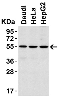
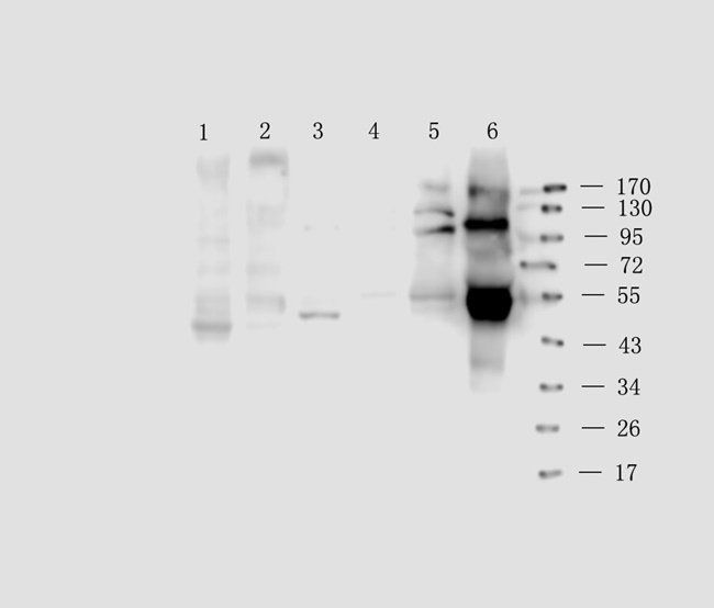
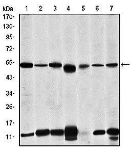
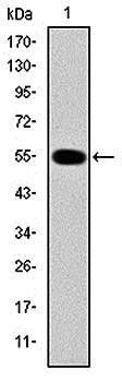
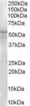
Western Blot Validation in Human Cell Lines. Loading: 15 µg/ of lysates per lane. Antibodies: IRAK4 orb1239512 (1 µg/mL), 1h incubation at RT in 5% NFDM/TBST. Secondary: Goat anti-rabbit IgG HRP conjugate at 1:10000 dilution.
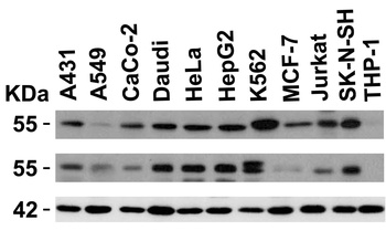
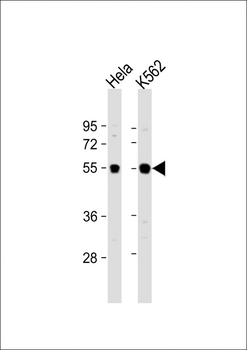
Independent Antibody Validation (IAV) via Protein Expression Profile in Cell Lines. Loading: 15 µg of lysates per lane. Antibodies: IRAK4 orb1239512 (1 µg/mL), IRAK4 orb1255476 (1 µg/mL), beta-actin (1.5 µg/mL), 1h incubation at RT in 5% NFDM/TBST. Secondary: Goat anti-rabbit IgG HRP conjugate at 1:10000 dilution.
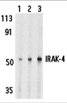
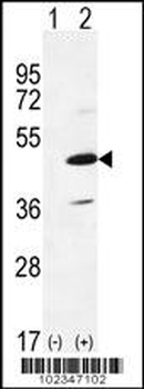
Western Blot Validation in Human HeLa Cell Lines. Loading: 15 µg of lysates per lane. Antibodies: IRAK4 orb1239512, 1h incubation at RT in 5% NFDM/TBST. Secondary: Goat anti-rabbit IgG HRP conjugate at 1:10000 dilution. Lane 1: 1 µg/mL, Lane 2: 2 µg/mL, Lane 3: 4 µg/mL.
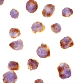
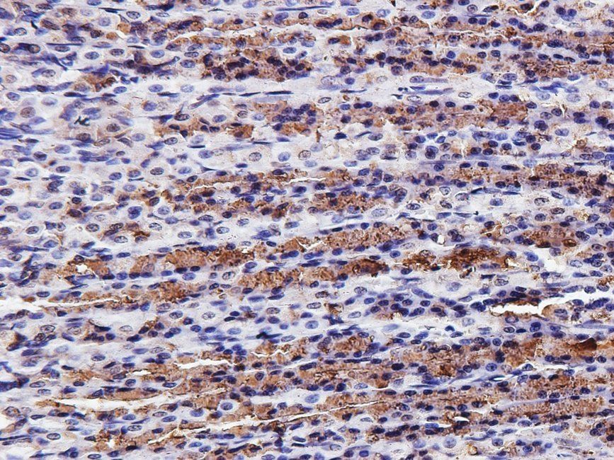
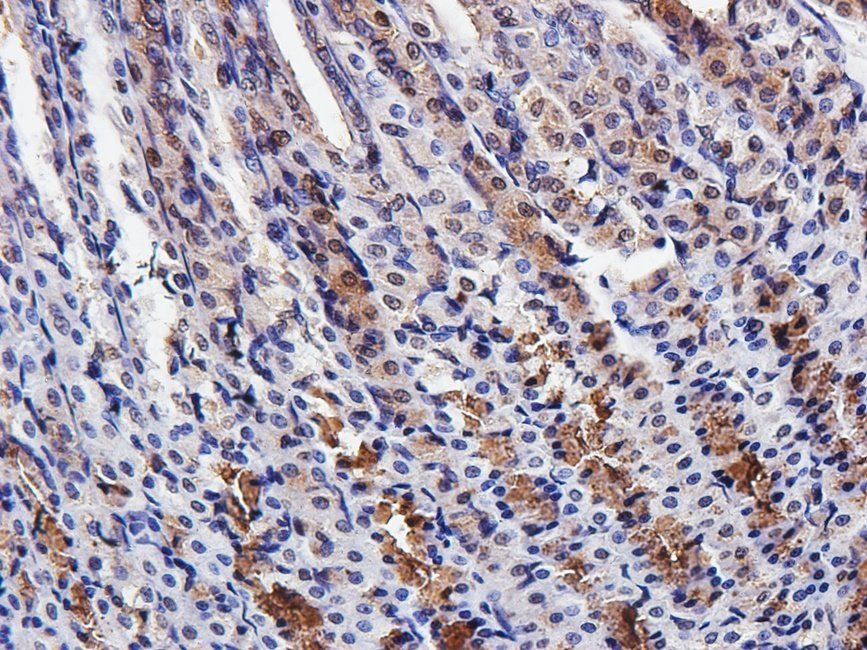
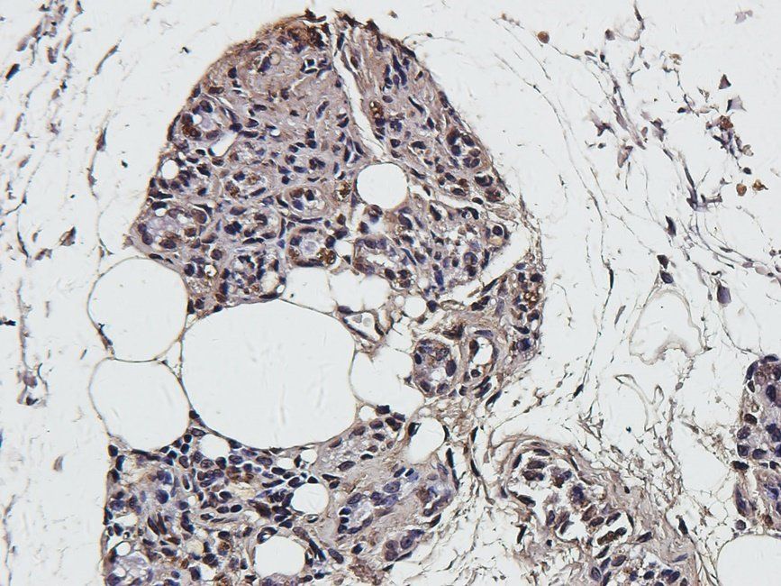
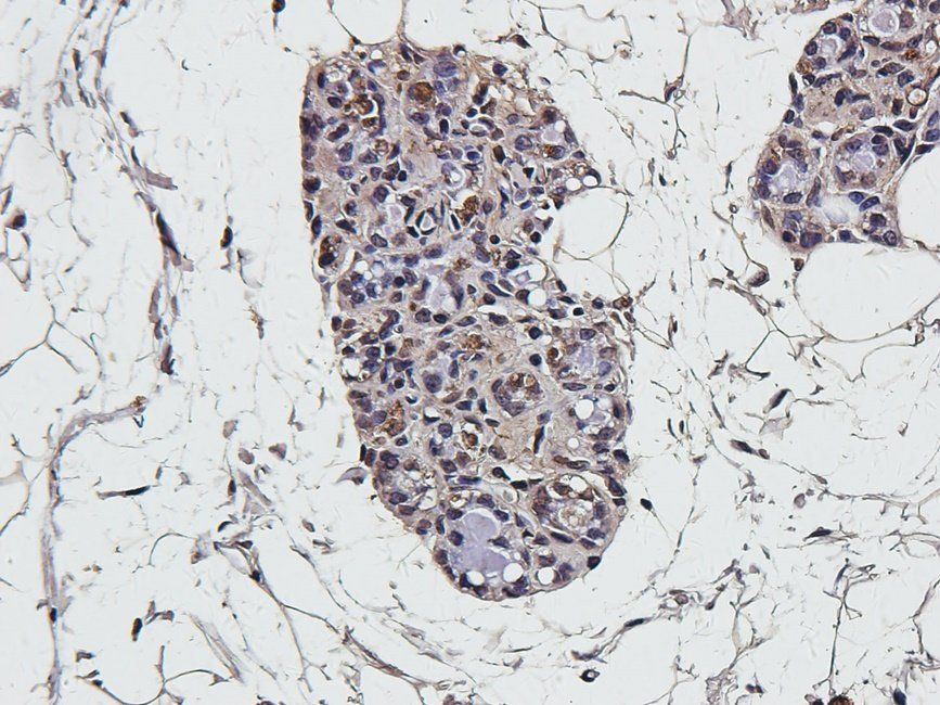
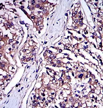
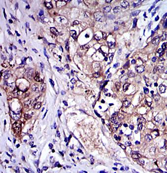
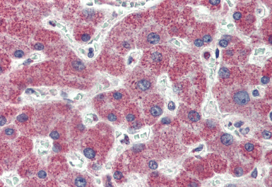
Immunocytochemistry Validation of IRAK4 in K562 Cells. Immunocytochemical analysis of K562 cells using anti-IRAK4 antibody (orb1239512) at 10 µg/ml. Cells was fixed with formaldehyde and blocked with 10% serum for 1 h at RT; antigen retrieval was by heat mediation with a citrate buffer (pH6). Samples were incubated with primary antibody overnight at 4°C. A goat anti-rabbit IgG H&L (HRP) at 1/250 was used as secondary. Counter stained with Hematoxylin.
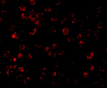
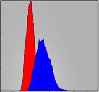
Immunofluorescence Validation of IRAK4 in K562 Cells. Immunofluorescent analysis of 4% paraformaldehyde-fixed K562 Cells labeling IRAK4 with orb1239512 at 10 µg/mL, followed by goat anti-rabbit IgG secondary antibody at 1/500 dilution (red).
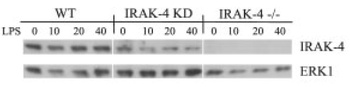
KO and KD Validation of IRAK4 in Mouse Bone Marrow-derived Macrophages (BMDM) (Koziczak-Holbro et al., 2008). Western blot analysis with anti-IRAK4 antibodies was performed for IRAK4 in BMDM isolated from the mice. IRAK4 expression was not observed in the IRAK-4-/- cells and also reduced in IRAK4 mutant (IRAK-4 KD) when compared with WT.
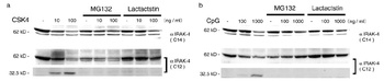
Regulated Expression Validation of IRAK4 in Mouse RAW 264 Cells (Hatao et al., 2004). RAW 264 cells were stimulated with (a) CSK4 and (b) CpG-DNA in the absence or presence of proteasome inhibitors (MG132 and Lactactstin). When detected with anti-IRAK4 antibodies (C12), IRAK4 expression was found to be reduced without the inhibitors. The smaller protein band, a cleavage product of IRAK4, was present in the absence of both inhibitors.
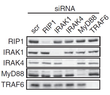
KD Validation of IRAK4 in HEK293T Cells (Heinz et al., 2012). Western blot analysis with anti-IRAK4 antibodies was performed for IRAK4 in HEK293T cells. IRAK4 expression was not observed in IRAK4 knockdown cells.
Documents Download
Datasheet
Product Information
Request a Document
Protocol Information
WB
Western Blot (IB, immunoblot)
IF
Immunofluorescence
ICC
Immunocytochemistry
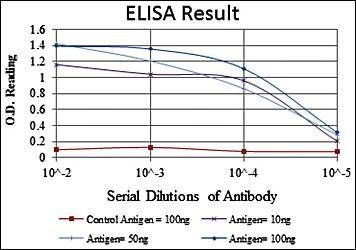
ELISA
Enzyme-linked Immunosorbent Assay (EIA)
IRAK4 Antibody (orb1239512)

- 0.0
Based on 0 reviews
Participating in our Biorbyt product reviews program enables you to support fellow scientists by sharing your firsthand experience with our products.
Login to Submit a ReviewAvailable Sizes
Select a size below
Free Secondary Antibody (20 ul)0/0
Please add an antibody product to your cart first.