You have no items in your shopping cart.
Interferon-inducible protein (IFITM3) Antibody (N-term)
SKU: orb1938125
Description
Research Area
Infectious Disease & Virology; Immunology & Inflammation
Images & Validation
−Item 1 of 6
| Tested Applications | IF, IHC-P, WB |
|---|---|
| Dilution Range | IF - 1:100-500, WB - 1:2000, IHC-P-Leica - 1:100 |
| Reactivity | Human, Mouse |
Key Properties
−| Host | Rabbit |
|---|---|
| Clonality | Polyclonal |
| Isotype | Rabbit IgG |
| Immunogen | This Interferon-inducible protein (IFITM3) antibody is generated from rabbits immunized with a KLH conjugated synthetic peptide between 1-30 amino acids from the N-terminal region of human Interferon-inducible protein (IFITM3). Antigen Region: 1-30 aa. |
| Target | IFITM3 (HGNC:5414) |
| Molecular Weight | 14632 Da |
| Conjugation | Unconjugated |
Storage & Handling
−| Storage | Maintain refrigerated at 2-8°C for up to 2 weeks. For long term storage store at -20°C in small aliquots to prevent freeze-thaw cycles |
|---|---|
| Form/Appearance | Purified polyclonal antibody supplied in PBS with 0.09% (W/V) sodium azide. This antibody is purified through a protein A column, followed by peptide affinity purification. |
| Expiration Date | 12 months from date of receipt. |
| Disclaimer | For research use only |
Alternative Names
−Interferon-induced transmembrane protein 3, Dispanin subfamily A member 2b, DSPA2b, Interferon-inducible protein 1-8U, IFITM3
Similar Products
−- Item 1 of 3
IFITM3 Rabbit Polyclonal Antibody [orb214984]
IF, IHC, WB
Human, Mouse, Porcine, Rat
Rabbit
Polyclonal
Unconjugated
30 μl, 100 μl, 200 μl, 50 μl - Item 1 of 1
- Item 1 of 1
IFITM3 Mouse Monoclonal Antibody [orb1473814]
WB
Human
Mouse
Monoclonal
Unconjugated
200 μl, 100 μl, 50 μl, 30 μl
Quality Guarantee
Explore bioreagents carefree to elevate your research. All our products are rigorously tested for performance. If a product does not perform as described on its datasheet, our scientific support team will provide expert troubleshooting, a prompt replacement, or a refund. For full details, please see our Terms & Conditions and Buying Guide. Contact us at [email protected].
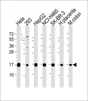
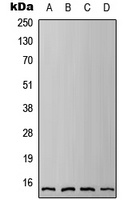
All lanes: Anti-IFITM3 Antibody (N-term) at 1:2000 dilution. Lane 1: Hela whole cell lysate. Lane 2: 293 whole cell lysate. Lane 3: HepG2 whole cell lysate. Lane 4: NCI-H460 whole cell lysate. Lane 5: SK-BR-3 whole cell lysate. Lane 6: human placenta lysate. Lane 7: mouse colon lysate.Lysates/proteins at 20 µg per lane. Secondary Goat Anti-Rabbit IgG, (H+L), Peroxidase conjugated at 1/10000 dilution. Predicted band size: 15 kDa. Blocking/Dilution buffer: 5% NFDM/TBST.
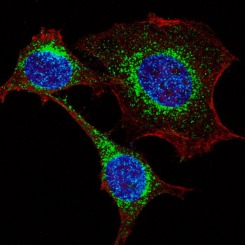
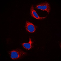
Fluorescent confocal image of HeLa cells stained with IFITM3 (N-term) Antibody. HeLa cells were fixed with 4% PFA (20 min), permeabilized with Triton X-100 (0.2%, 30 min), then incubated with Fragilis (IFITM3) (N-term) primary antibody (1:200, 2 h at room temperature). For secondary antibody, Alexa Fluor 488 conjugated donkey anti-rabbit antibody (green) was used (1:1000, 1h). Cytoplasmic actin was counterstained with Alexa Fluor 555 (red) conjugated Phalloidin (5.25 μM, 25 min). Nuclei were counterstained with Hoechst 33342 (blue) (10 µg/ml, 3 min).
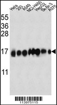
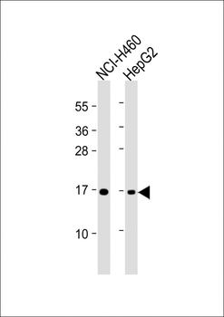
IFITM3 Antibody (N-term) western blot analysis in Hela, 293, A549, NCI-H460, HepG2, Sk-Br-3, A375 cell line lysates (35 ug/lane). This demonstrates the IFITM3 antibody detected the IFITM3 protein (arrow).
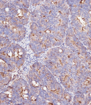
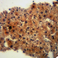
Immunohistochemical analysis on paraffin-embedded Human colon carcinoma tissue was performed on the Leica BOND RXm. Tissue was fixed with formaldehyde at room temperature. Heat induced epitope retrieval was performed by EDTA buffer (pH9.0). Samples were incubated with primary antibody (1:100) for 15min at room temperature. Leica Bond Polymer Refine Detection was used as the secondary Antibody.
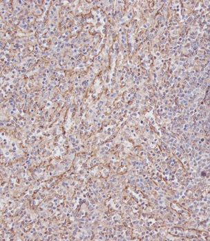
Immunohistochemical analysis on paraffin-embedded Human spleen tissue was performed on the Leica BOND RXm. Tissue was fixed with formaldehyde at room temperature. Heat induced epitope retrieval was performed by EDTA buffer (pH9.0). Samples were incubated with primary antibody (1:100) for 15min at room temperature. Leica Bond Polymer Refine Detection was used as the secondary Antibody.
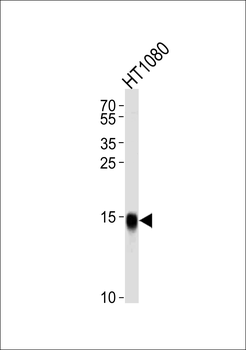
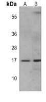
Western blot analysis of lysate from HT1080 cell line, using IFITM3 Antibody (N-term).diluted at 1:1000 at each lane. A goat anti-rabbit IgG H&L (HRP) at 1:5000 dilution was used as the secondary Antibody. Lysate at 35 ug per lane.
Quick Database Links
UniProt Details
− No UniProt data available
NCBI Reference Sequences
−Associated Accession Numbers
Curated reference sequences for the gene transcript and protein product| Protein | NP_066362.2 |
|---|
Documents Download
Datasheet
Product Information
Request a Document
Protocol Information
WB
Western Blot (IB, immunoblot)
IHC-P
Immunohistochemistry Paraffin
IF
Immunofluorescence
Interferon-inducible protein (IFITM3) Antibody (N-term) (orb1938125)

- 0.0
Based on 0 reviews
Participating in our Biorbyt product reviews program enables you to support fellow scientists by sharing your firsthand experience with our products.
Login to Submit a ReviewAvailable Sizes
Select a size below
Choose Conjugation or Carrier Free Version
Free Secondary Antibody (20 ul)0/0
Please add an antibody product to your cart first.