You have no items in your shopping cart.
Featured
Description
Research Area
Metabolism Research
Images & Validation
−Item 1 of 5
| Tested Applications | IF, IHC-Fr, IHC-P |
|---|---|
| Dilution Range | IHC-P=1:50-200, IHC-F=1:50-200, IF=1:50-200 |
| Reactivity | Human |
Key Properties
−| Antibody Type | Primary Antibody |
|---|---|
| Host | Rabbit |
| Clonality | Recombinant |
| Isotype | IgG |
| Immunogen | Recombinant human IGF2 protein (25-121 /180aa) |
| Target | IGF2 |
| Molecular Weight | 11 kDa |
| Purification | Affinity purified by Protein A |
| Conjugation | Unconjugated |
Storage & Handling
−| Storage | Maintain refrigerated at 2-8°C for up to 2 weeks. For long term storage store at -20°C in small aliquots to prevent freeze-thaw cycles. |
|---|---|
| Form/Appearance | Liquid |
| Buffer/Preservatives | 0.01M TBS (pH7.4) with 1% rAlbumin, 0.02% Proclin300 and 50% Glycerol. |
| Concentration | 1mg/ml |
| Expiration Date | 12 months from date of receipt. |
| Disclaimer | For research use only |
Alternative Names
−C11orf43; GRDF; IGF-II; PP9974; SRS3; Igf-2; M6pr; Mpr; Peg2; IGFII; RNIGF2; IGF2_BOVIN; IGF2; Erythrotropin; Insulin-like growth factor II (IGF-II); IGF2_HUMAN; Somatomedin-A; T3M-11-derived growth factor; IGF2_MOUSE; Multiplication-stimulating polypeptide; IGF2_RAT; Multiplication-stimulating activity (MSA); insulin like growth factor 2; chromosome 11 open reading frame 43; insulin-like growth factor 2; somatomedin A; preptin
Similar Products
−- Item 1 of 3
Recombinant Glypican 3 Antibody / GPC3 / Rabbit Monoclonal [orb534018]
FACS, IF, IHC-P
Human
Rabbit
Recombinant
Unconjugated
100 μg, 20 μg - Item 1 of 3
Recombinant Glypican 3 Antibody / GPC3 / Rabbit Monoclonal [orb2640955]
FACS, IF, IHC-P
Human
Rabbit
Recombinant
Unconjugated
100 μg - Item 1 of 1
IGF2R/M6PR Recombinant Rabbit Monoclonal Antibody [orb1974640]
WB
Mouse, Rat
Human
Rabbit
Recombinant
Unconjugated
50 μl, 100 μl, 25 μl - Item 1 of 1
IGF2BP2 Recombinant Rabbit Monoclonal Antibody [orb2561452]
FC, IF, IHC-Fr, IHC-P, KO/KD Validated, WB
Human, Mouse, Rat
Human, Mouse, Rat
Rabbit
Recombinant
Unconjugated
50 μl, 100 μl, 25 μl 
IGF2 Rabbit Monoclonal Antibody [orb3072877]
IF, WB
Human, Rat
Rabbit
Monoclonal
Unconjugated
200 μl, 100 μl, 50 μl, 30 μl


Quality Guarantee
Explore bioreagents carefree to elevate your research. All our products are rigorously tested for performance. If a product does not perform as described on its datasheet, our scientific support team will provide expert troubleshooting, a prompt replacement, or a refund. For full details, please see our Terms & Conditions and Buying Guide. Contact us at [email protected].
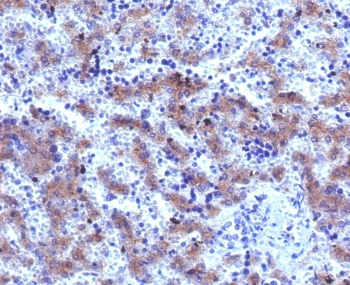
Immunohistochemical analysis of paraffin-embedded human breast carcinoma tissue using anti-IGF2 antibody. The section was pre-treated using heat mediated antigen retrieval with Tris-EDTA buffer (pH9.0) for 20 minutes. The tissues were blocked in 1% BSA for 30 minutes at room temperature, washed with ddH2O and PBS, and then probed with the primary antibody (orb1499377, 1/50) for 30 minutes at room temperature. The detection was performed using an HRP conjugated compact polymer system. DAB was used as the chromogen. Tissues were counterstained with hematoxylin and mounted with DPX.
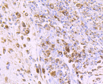
Immunohistochemical analysis of paraffin-embedded human colon carcinoma tissue using anti-IGF2 antibody. The section was pre-treated using heat mediated antigen retrieval with Tris-EDTA buffer (pH9.0) for 20 minutes. The tissues were blocked in 1% BSA for 30 minutes at room temperature, washed with ddH2O and PBS, and then probed with the primary antibody (orb1499377, 1/50) for 30 minutes at room temperature. The detection was performed using an HRP conjugated compact polymer system. DAB was used as the chromogen. Tissues were counterstained with hematoxylin and mounted with DPX.
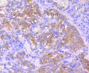
Immunohistochemical analysis of paraffin-embedded human liver carcinoma tissue using anti-IGF2 antibody. The section was pre-treated using heat mediated antigen retrieval with Tris-EDTA buffer (pH9.0) for 20 minutes. The tissues were blocked in 1% BSA for 30 minutes at room temperature, washed with ddH2O and PBS, and then probed with the primary antibody (orb1499377, 1/50) for 30 minutes at room temperature. The detection was performed using an HRP conjugated compact polymer system. DAB was used as the chromogen. Tissues were counterstained with hematoxylin and mounted with DPX.
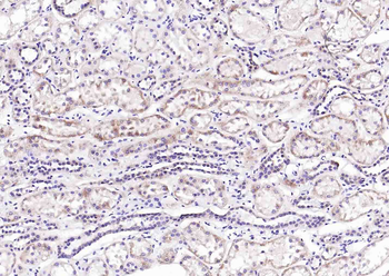
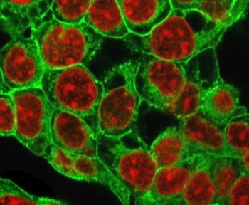
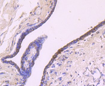
Paraformaldehyde-fixed, paraffin embedded (Human kidney), Antigen retrieval by boiling in sodium citrate buffer (pH6.0) for 15 min, Block endogenous peroxidase by 3% hydrogen peroxide for 20 minutes, Blocking buffer (normal goat serum) at 37°C for 30 min, Antibody incubation with (IGF2) Monoclonal Antibody, Unconjugated (orb1499377) at 1:200 overnight at 4°C, followed by operating according to SP Kit (Rabbit) instructionsand DAB staining.
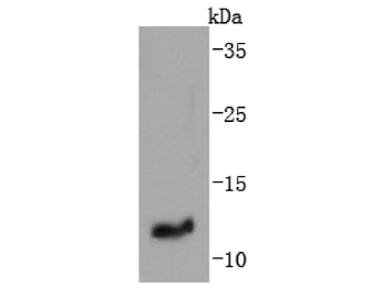
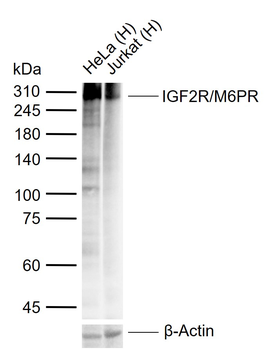
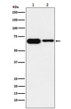
Western blot analysis of IGF2 on human placenta tissue lysates. Proteins were transferred to a PVDF membrane and blocked with 5% BSA in PBS for 1 hour at room temperature. The primary antibody (orb1499377, 1/500) was used in 5% BSA at room temperature for 2 hours. Goat Anti-Rabbit IgG - HRP Secondary Antibody (HA1001) at 1:200000 dilution was used for 1 hour at room temperature. Predicted band size: 20 kDa, Observed band size: 11 kDa.
Quick Database Links
Gene Symbol
IGF2
UniProt
UniProt Details
− No UniProt data available
Documents Download
Datasheet
Product Information
Request a Document
Protocol Information
IHC-P
Immunohistochemistry Paraffin
IHC-Fr
Immunohistochemistry Frozen
IF
Immunofluorescence
IGF2 Recombinant Rabbit Monoclonal Antibody (orb1499377)

- 0.0
Based on 0 reviews
Participating in our Biorbyt product reviews program enables you to support fellow scientists by sharing your firsthand experience with our products.
Login to Submit a ReviewAvailable Sizes
Select a size below
Free Secondary Antibody (20 ul)0/0
Please add an antibody product to your cart first.