You have no items in your shopping cart.
Description
Research Area
Metabolism Research
Images & Validation
−Item 1 of 6
| Tested Applications | ELISA, FC, IF, IHC, IP, Multiplex Assay, WB |
|---|---|
| Dilution Range | ELISA: 1:5000-1:50000, FC: 0.5-1x10^6 cells, IF: 1:50-1:100, IP: 10-100 µL, WB: 1:500-1:1500 |
| Reactivity | Mouse |
| Application Notes |
Key Properties
−| Antibody Type | Primary Antibody |
|---|---|
| Host | Mouse |
| Clonality | Monoclonal |
| Isotype | IgG1 |
| Clone No. | 2E2.6 |
| Immunogen | IDO1 antibody was produced in mouse by repeated immunizations with mouse recombinant IDO1 protein followed by hybridoma development. |
| Target | Ido1 |
| Purity | Anti-IDO1 antibody was purified from concentrated tissue culture supernate by Protein G chromatography followed by extensive dialysis against the buffer stated above. IDO1 antibody is specific for mouse IDO1 protein. Mouse IDO1 does not react with human tissues. Cross-reactivity with IDO1 from other sources has not been determined. |
| Conjugation | Unconjugated |
Storage & Handling
−| Storage | Store vial at -20° C or below prior to opening. This vial contains a relatively low volume of reagent (25 µL). To minimize loss of volume dilute 1:10 by adding 225 µL of the buffer stated above directly to the vial. Recap, mix thoroughly and briefly centrifuge to collect the volume at the bottom of the vial. Use this intermediate dilution when calculating final dilutions as recommended below. Store the vial at -20°C or below after dilution. Avoid cycles of freezing and thawing. |
|---|---|
| Form/Appearance | Liquid (sterile filtered) |
| Buffer/Preservatives | Preservative: 0.01% (w/v) Sodium Azide. Stabilizer: None; Buffer: 0.02 M Potassium Phosphate, 0.15 M Sodium Chloride, pH 7.2 |
| Concentration | 0.95 mg/mL |
| Expiration Date | 12 months from date of receipt. |
| Dry Ice Shipping | Please note: This product requires shipment on dry ice. A dry ice surcharge will apply. |
| Disclaimer | For research use only |
Alternative Names
−mouse anti-IDO1 antibody, Ido, Indo, Indoleamine 2,3-dioxygenase 1, Indoleamine-pyrrole 2,3-dioxygenase, Ido 1, Ido-1, IDO1 antibody, mouse anti-IDO-1 antibody
Similar Products
−- Item 1 of 9
IDO Rabbit Polyclonal Antibody [orb157614]
ICC, IF, IHC-P, WB
Guinea pig, Human, Mouse, Rat
Rabbit
Polyclonal
Unconjugated
100 μg - Item 1 of 4
IDO1 Rabbit Polyclonal Antibody [orb500950]
WB
Bovine, Canine, Equine, Porcine, Rabbit, Rat, Sheep
Human, Mouse, Rat
Rabbit
Polyclonal
Unconjugated
50 μl, 100 μl, 200 μl - Item 1 of 5
- Item 1 of 6
Ido1 Antibody [orb345210]
ELISA, FC, IF, IHC, IP, Multiplex Assay, WB
Mouse
Mouse
Monoclonal
Unconjugated
100 μg - Item 1 of 4
Indoleamine 2, 3-dioxygenase/IDO1 Rabbit Polyclonal Antibody [orb312129]
FC, ICC, IF, IHC, WB
Human
Rabbit
Polyclonal
Unconjugated
100 μg


Quality Guarantee
Explore bioreagents carefree to elevate your research. All our products are rigorously tested for performance. If a product does not perform as described on its datasheet, our scientific support team will provide expert troubleshooting, a prompt replacement, or a refund. For full details, please see our Terms & Conditions and Buying Guide. Contact us at [email protected].

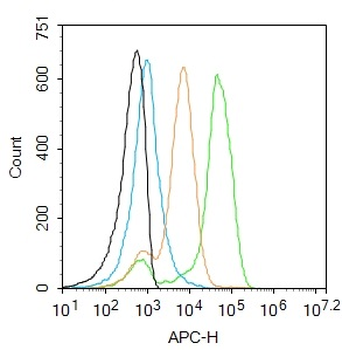
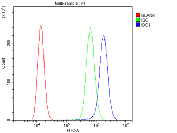
Flow Cytometry of Mouse Anti-IDO1 antibody. Cells: HEK293 cells. Expresing: mouse IDO-1(blue) and mouse IDO-2 (red). Primary antibody: IDO1 (2E2) monoclonal antibody. Secondary antibody: Biotin mouse secondary antibody at 1:10000 for 45 min at RT and streptavidin PE at 1:5000 for 30 min at RT.
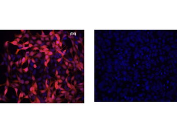
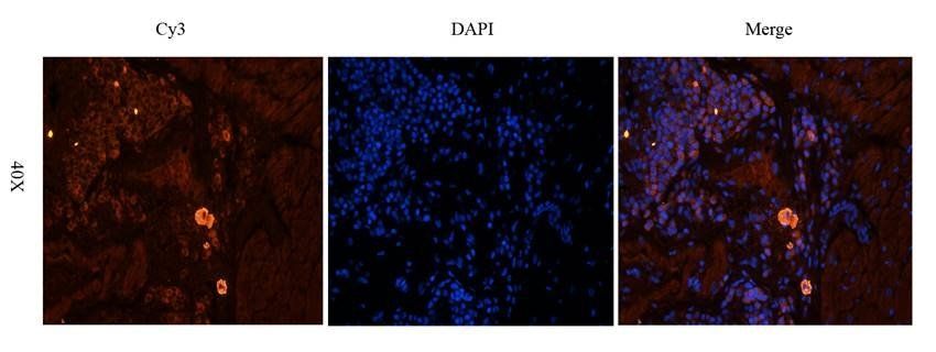
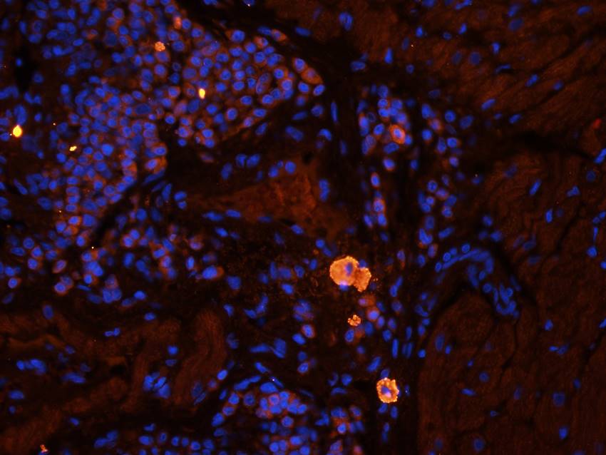
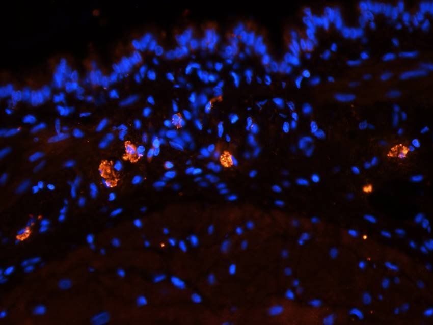
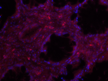
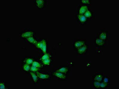
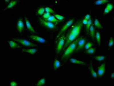
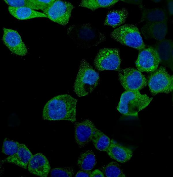
Immunofluorescence Microscopy of Mouse Anti-IDO1 Antibody. Cells: HEK293 cells. Fixation: 0.5% PFA. Expressing: mouse IDO-1 (left) and mouse IDO-2 (right). Primary antibody: IDO1 (2E2) monoclonal antibody. Antigen retrieval: not required. Secondary antibody: mouse secondary antibody at 1:10000 for 45 min at RT. Localization: IDO-1 is located in the cytosol. Staining: IDO1 as red fluorescent signal with bis-benzimide nuclear counterstain (blue).

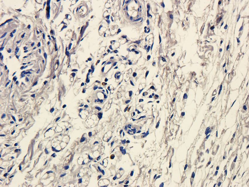
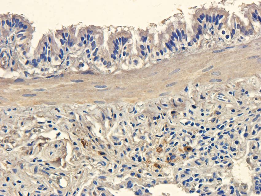
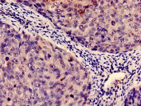
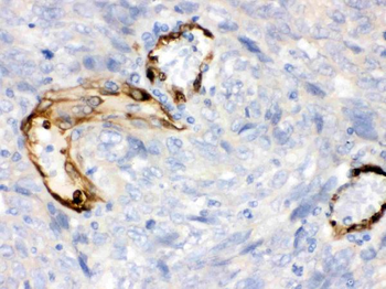
Immunohistochemistry of Mouse Anti-IDO1 Antibody. Tissue: epididymis from wild-type (left) or IDO1 null mice (right). Fixation: frozen sections. Antigen retrieval: not required. Primary antibody: IDO1 (2E2) monoclonal antibody. Secondary antibody: Peroxidase mouse secondary antibody at 1:10000 for 45 min at RT. Localization: IDO-1 is located in the cytosol. Staining: IDO 1 as precipitated brown signal.
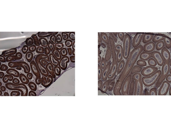
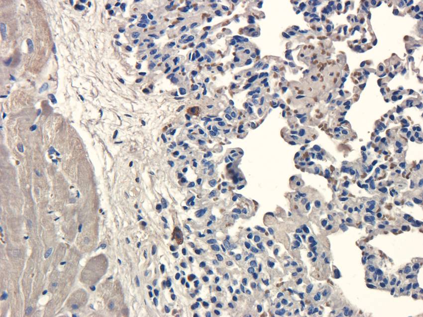
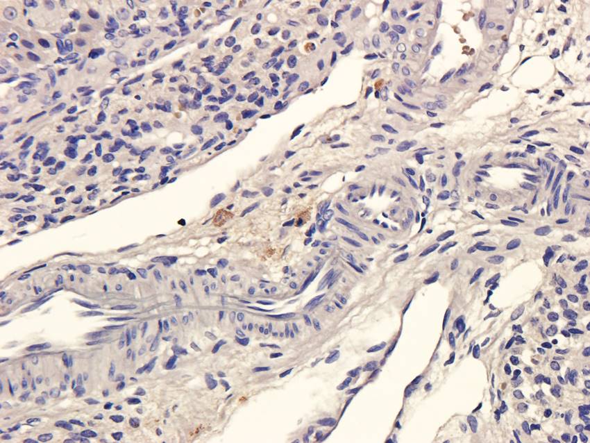
Immunohistochemistry of Mouse anti-IDO1 antibody. Tissue: epididymis from wild-type (left) or IDO1 null mice (right). Fixation: paraffin-embedded. Primary antibody: IDO1 (2E2) monoclonal antibody. Secondary antibody: Peroxidase mouse secondary antibody at 1:10000 for 45 min at RT. Localization: IDO-1 is located in the cytosol. Staining: IDO 1 as precipitated brown signal.
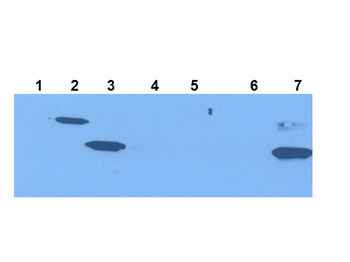
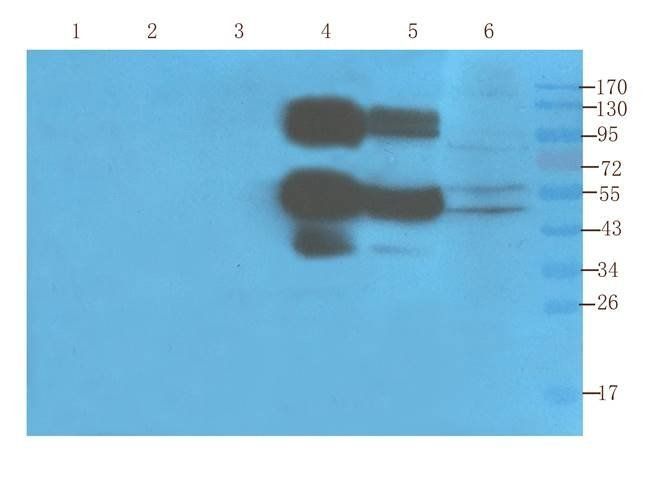
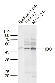
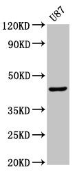
Western Blot of Mouse Anti-IDO1 Antibody. Extracts from 293HEK Cells expressing: Lane 1: Control Vector. Lane 2: His-tagged mouse IDO1. Lane 3: mouse IDO1. Lane 4: His-tagged mouse IDO2. Lane 5: mouse IDO2. Lane 6: Epididymis from IDO null. Lane 7: wild type mice. Primary antibody: IDO-1(2E2) monoclonal antibody. Secondary antibody: IRDye800™ mouse secondary antibody at 1:10000 for 45 min at RT. Block: 1xPBST overnight at 4°C. Predicted/Observed size: 41-42 kDa/41-42 kDa for IDO-1. Other band(s): none.

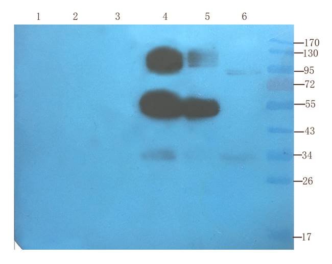
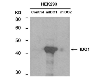
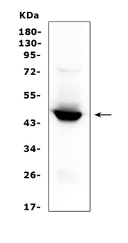
Western Blot of mouse anti-IDO1 antibody. Lane 1: HEK293 control vector. Lane 2: HEK293 expressing mouse IDO1. Lane 3: HEK293 expressing mouse IDO2. Load: 35 µg per lane. Primary antibody: IDO 1 antibody at 1:400 for overnight at 4°C. Secondary antibody: IRDye800™ mouse secondary antibody at 1:10000 for 45 min at RT. Block: 5% BLOTTO overnight at 4°C. Predicted/Observed size: 45.6 kDa, ~44 kDa for IDO1. Other band(s): non-specifics.
Quick Database Links
UniProt Details
− No UniProt data available
NCBI Reference Sequences
−Associated Accession Numbers
Curated reference sequences for the gene transcript and protein product| Protein | NP_032350.1 |
|---|
Documents Download
Datasheet
Product Information
Request a Document
Protocol Information
WB
Western Blot (IB, immunoblot)
IHC
Immunohistochemistry
FC
Flow Cytometry
IF
Immunofluorescence
ELISA
Enzyme-linked Immunosorbent Assay (EIA)
IP
Immunoprecipitation
Ido1 Antibody (orb345211)

- 0.0
Based on 0 reviews
Participating in our Biorbyt product reviews program enables you to support fellow scientists by sharing your firsthand experience with our products.
Login to Submit a ReviewAvailable Sizes
Select a size below
Free Secondary Antibody (20 ul)0/0
Please add an antibody product to your cart first.