You have no items in your shopping cart.
Description
Research Area
Protein Biochemistry
Images & Validation
−Item 1 of 3
| Tested Applications | DOT, ELISA, IHC, WB |
|---|---|
| Application Notes |
Key Properties
−| Source | Human |
|---|---|
| Biological Origin | Human |
| Target | TF |
| Isotype | Transferrin |
| Conjugation | HRP |
| Purity | Human Transferrin Peroxidase conjugated was prepared from normal serum by a multi-step process including selective precipitation and tandem chromatography followed by extensive dialysis against the buffer stated above. Human Transferrin Peroxidase conjugated was assayed by immunoelectrophoresis and resulted in a single precipitin arc against anti-Peroxidase, anti-Human Transferrin and anti-Human Serum. |
Storage & Handling
−| Storage | Store vial at 4° C prior to restoration. For extended storage aliquot contents and freeze at -20° C or below. Avoid cycles of freezing and thawing. Centrifuge product if not completely clear after standing at room temperature. Human Transferrin Peroxidase conjugated is stable for several weeks at 4° C as an undiluted liquid. Dilute only prior to immediate use. |
|---|---|
| Form/Appearance | Lyophilized |
| Buffer/Preservatives | Preservative: 0.01% (w/v) Gentamicin Sulfate. Do NOT add Sodium Azide!. Stabilizer: 10 mg/mL Bovine Serum Albumin (rAlbumin) - Immunoglobulin and Protease free; Buffer: 0.02 M Potassium Phosphate, 0.15 M Sodium Chloride, pH 7.2 |
| Concentration | 1.0 mg/ml |
| Expiration Date | 6 months from date of receipt. |
| Disclaimer | For research use only |
Alternative Names
−Human transferrin peroxidase conjugation, HRP conjugated transferrin
Similar Products
−- Item 1 of 2
Recombinant Eosinophil Peroxidase Antibody [orb699615]
FACS, IF, IHC-P
Human
Mouse
Recombinant
Unconjugated
100 μg, 20 μg - Item 1 of 2
Recombinant Eosinophil Peroxidase Antibody [orb2644492]
FACS, IF, IHC-P
Human
Mouse
Recombinant
Unconjugated
100 μg - Item 1 of 2
Recombinant EPX Antibody / Eosinophil Peroxidase [orb2644494]
FACS, IF, IHC-P
Human
Rabbit
Recombinant
Unconjugated
100 μg - Item 1 of 2
Recombinant EPX Antibody / Eosinophil Peroxidase [orb699616]
FACS, IF, IHC-P
Human
Rabbit
Recombinant
Unconjugated
100 μg, 20 μg 
Eosinophil Peroxidase Antibody / EPO [orb749398]
ELISA, FACS, IF
Human
Mouse
Monoclonal
Unconjugated
100 μg, 20 μg


Quality Guarantee
Explore bioreagents carefree to elevate your research. All our products are rigorously tested for performance. If a product does not perform as described on its datasheet, our scientific support team will provide expert troubleshooting, a prompt replacement, or a refund. For full details, please see our Terms & Conditions and Buying Guide. Contact us at [email protected].
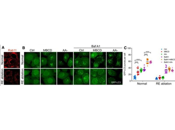
CEMM disruption releases VAMP3 from CEMMs at recycling endosomal membrane. Used a recycling endosome ablation approach by using combined treatment with 3, 3'-diaminobenzidine (DAB) and H2O2 to the cells pre-loaded with horseradish peroxidase-transferrin (HRP-TF).60, 61 The significant reduction of the RAB11 signaling in the recycling endosome-ablated cells (RE ablation) proved the ablation efficiency of this method (Figure 3A). Importantly, CEMM disruption-induced autophagic flux is almost totally blocked by RE ablation (Figures 3B and 3C), indicating the importance of recycling endosomes in CEMM disruption-induced autophagosome formation (A) After ablation of recycling endosomes, HeLa cells were immunostained with Rab11 (red), and observed under a confocal microscope (×600). Scale bars, 5 µm. (B) After ablation of recycling endosome, HeLa cells with stable expression of GFP-LC3B were pre-treated with MBCD (5 mM, 1 h) and then incubated in the presence or absence of Baf A1 (100 nM). Then cells were observed under a confocal microscope (×600). Scale bars, 5 µm. (C) The number of GFP-LC3 puncta observed in (B) are presented as means ± SD. ∗∗∗∗p < 0.0001.
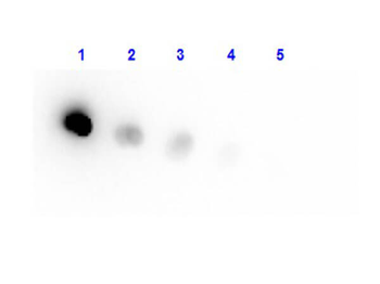
Dot Blot result of Human Transferrin Peroxidase conjugate. Dots are Human Transferrin HRP at (1) 100 ng, (2) 33.3 ng, (3) 11.1 ng, (4) 3.70 ng, (5) 1.23 ng. Blocking: orb348637 for 60 min at RT. Primary Antibody: none. Secondary Antibody: none.
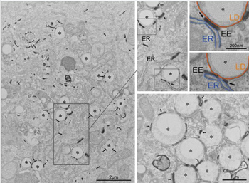
The figure illustrates that EE tubules, defined by the presence of internalized transferrin, interact with LDs in mammalian cells. BHK cells were cotransfected with a plasmid encoding human transferrin receptor (TfR plus plasmid encoding GFP) and then incubated with transferrin-HRP for 30 min at 37°C. Cells were then processed for HRP detection and processed for electron microscopy using a mild fixation/low membrane-contrast staining method to optimize transferrin-HRP visualization. Transferrin-HRP-labeled EE tubules (arrows) were specifically associated with LDs (asterisks) as shown in the low magnification overview (left panel) and at higher magnification in the lower right panel. The two pseudocolored panels show higher magnification views of the neighboring panels, with ER in blue and the LD monolayer in orange; note the tripartite interaction with the EE tubule (EE, arrows).
Quick Database Links
UniProt Details
− No UniProt data available
NCBI Reference Sequences
−Associated Accession Numbers
Curated reference sequences for the gene transcript and protein product| RefSeq | AAB22049.1 |
|---|
Documents Download
Datasheet
Product Information
Request a Document
Protocol Information
Protein Handling and Storage Guide
Protein Handling Guide
WB
Western Blot (IB, immunoblot)
IHC
Immunohistochemistry
ELISA
Enzyme-linked Immunosorbent Assay (EIA)
DOT
Dot Blot
Human Transferrin protein (Peroxidase) (orb346250)

- 0.0
Based on 0 reviews
Participating in our Biorbyt product reviews program enables you to support fellow scientists by sharing your firsthand experience with our products.
Login to Submit a ReviewAvailable Sizes
Select a size below