You have no items in your shopping cart.
Description
Research Area
Images & Validation
−| Tested Applications | ELISA, FC, WB |
|---|---|
| Reactivity | Human |
Key Properties
−| Antibody Type | Recombinant Antibody |
|---|---|
| Host | Mouse |
| Clonality | Monoclonal |
| Isotype | IgG2b-kappa |
| Clone No. | BDDOX3 |
| Purity | >95% as determined by SDS-PAGE. |
| Purification | Protein A/G purified from cell culture supernatant. |
| Endotoxin | Please contact with the lab for this information. |
| Conjugation | Unconjugated |
Storage & Handling
−| Storage | Maintain refrigerated at 2-8°C for up to 2 weeks. For long term storage store at -20°C in small aliquots to prevent freeze-thaw cycles. |
|---|---|
| Form/Appearance | Liquid |
| Buffer/Preservatives | 0.01M PBS, pH 7.4. |
| Expiration Date | 12 months from date of receipt. |
| Disclaimer | For research use only |
Alternative Names
−Similar Products
−Beta 2 Microglobulin B2M Mouse Monoclonal Antibody [orb507558]
FC, ICC, IF, IHC, WB
Human, Monkey
Mouse
Monoclonal
Unconjugated
100 μgBeta 2 Microglobulin Mouse Monoclonal Antibody [orb1600140]
WB
Human
Mouse
Monoclonal
Unconjugated
200 μg, 200 μl, 50 μl, 100 μlBeta 2 Microglobulin Mouse Monoclonal Antibody [orb2563589]
ELISA
Human
Human
Mouse
Monoclonal
Unconjugated
inquiryBeta 2 Microglobulin Mouse Monoclonal Antibody [orb2563578]
ELISA
Human
Human
Mouse
Monoclonal
Unconjugated
inquiry

Quality Guarantee
Explore bioreagents carefree to elevate your research. All our products are rigorously tested for performance. If a product does not perform as described on its datasheet, our scientific support team will provide expert troubleshooting, a prompt replacement, or a refund. For full details, please see our Terms & Conditions and Buying Guide. Contact us at [email protected].

Detects B2M/Beta-2-microglobulin in indirect ELISAs.

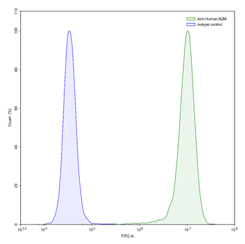
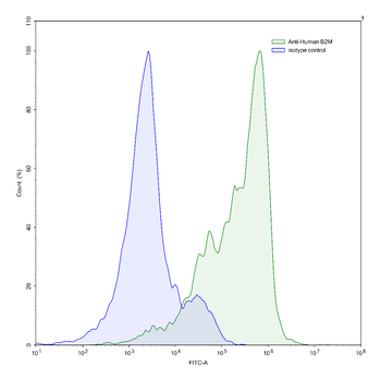
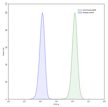
Flow-cytometry using anti-human B2M antibody. A431 cells were stained with an irrelevant antibody (Blue Histogram) or an anti-human B2M antibody monoclonal antibody (Catalog # orb2960486, Green Histogram) at a concentration of 5 µg/mL for 30 mins at RT. After washing, bound antibody was detected using a FITC conjugated goat anti-mouse antibody and cells analysed on a NovoCyte Flow Cytometer.

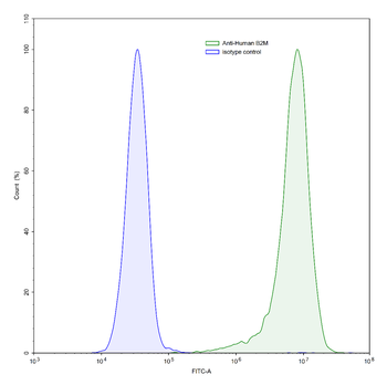
Flow-cytometry using anti-human B2M antibody. HeLa cells were stained with an irrelevant antibody (Blue Histogram) or an anti-human B2M antibody monoclonal antibody (Catalog # orb2960486, Green Histogram) at a concentration of 5 µg/mL for 30 mins at RT. After washing, bound antibody was detected using a FITC conjugated goat anti-mouse antibody and cells analysed on a NovoCyte Flow Cytometer.

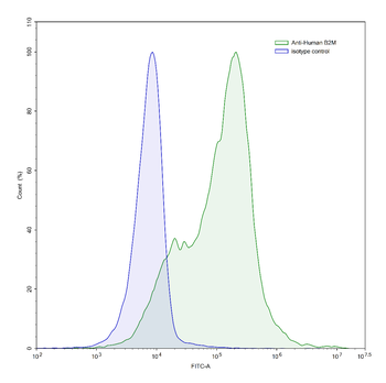
Flow-cytometry using anti-human B2M antibody. Human peripheral blood lymphocytes were stained with an irrelevant antibody (Blue Histogram) or an anti-human B2M antibody monoclonal antibody (Catalog # orb2960486, Green Histogram) at a concentration of 5 µg/mL for 30 mins at RT. After washing, bound antibody was detected using a FITC conjugated goat anti-mouse antibody and cells analysed on a NovoCyte Flow Cytometer.

Flow-cytometry using anti-human B2M antibody. Human peripheral blood lymphocytes were stained with an irrelevant antibody (Blue Histogram) or an anti-human B2M antibody monoclonal antibody (Catalog # orb2960486, Green Histogram) at a concentration of 5 µg/mL for 30 mins at RT. After washing, bound antibody was detected using a FITC conjugated goat anti-mouse antibody and cells analysed on a NovoCyte Flow Cytometer.

Flow-cytometry using anti-human B2M antibody. Jurkat cells were stained with an irrelevant antibody (Blue Histogram) or an anti-human B2M antibody monoclonal antibody (Catalog # orb2960486, Green Histogram) at a concentration of 5 µg/mL for 30 mins at RT. After washing, bound antibody was detected using a FITC conjugated goat anti-mouse antibody and cells analysed on a NovoCyte Flow Cytometer.

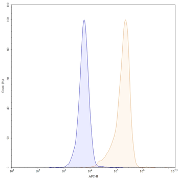
Flow-cytometry using APC anti-human B2M antibody. Hela cells were stained with an irrelevant antibody (Blue Histogram) or an APC anti-human B2M monoclonal antibody (Catalog: orb2960486, Yellow Histogram) at a concentration of 5 µg/ml for 30 mins at RT. After washing, and cells analysed on a NovoCyte Flow Cytometer.

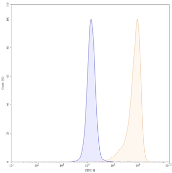
Flow-cytometry using FITC anti-human B2M antibody. Hela cells were stained with an irrelevant antibody (Blue Histogram) or an FITC anti-human B2M monoclonal antibody (Catalog: orb2960486, Yellow Histogram) at a concentration of 5 µg/ml for 30 mins at RT. After washing, and cells analysed on a NovoCyte Flow Cytometer.

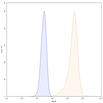
Flow-cytometry using PE anti-human B2M antibody. Hela cells were stained with an irrelevant antibody (Blue Histogram) or an PE anti-human B2M monoclonal antibody (Catalog: orb2960486, Yellow Histogram) at a concentration of 5 µg/ml for 30 mins at RT. After washing, and cells analysed on a NovoCyte Flow Cytometer.

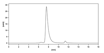
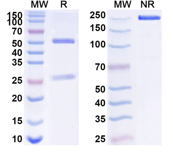
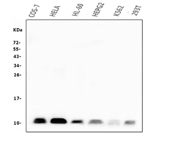
SDS-PAGE for Anti-Human B2M/Beta-2-microglobulin Antibody.

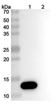
Western blot analysis was performed using anti-CD1a & B2M monoclonal antibody at 1 µg/mL on various samples. Lane 1: recombinant human CD1a & B2M (Catalog No: orb2966452), Lane 2: negative control.
Quick Database Links
UniProt Details
−Documents Download
Request a Document
Protocol Information
B2M/Beta-2-microglobulin Mouse Monoclonal Antibody (orb2960486)
Participating in our Biorbyt product reviews program enables you to support fellow scientists by sharing your firsthand experience with our products.
Login to Submit a Review


![Anti-Beta-2 microglobulin [BBM.1]](/images/pub/media/catalog/product/NewWebsite/35/orb613794_1.png)
![Anti-Beta-2 microglobulin [BBM.1]](/images/pub/media/catalog/product/NewWebsite/35/orb613794_2.png)
![Anti-Beta-2 microglobulin [BBM.1]](/images/pub/media/catalog/product/NewWebsite/35/orb613794_3.png)