You have no items in your shopping cart.
Featured
Description
Research Area
Cancer Biology, Cell Biology, Protein Biochemistry, Signal Transduction
Images & Validation
−Item 1 of 4
| Tested Applications | ELISA, ICC, IF, IHC, WB |
|---|---|
| Dilution Range | WB (1:1000), IHC (1:100), ICC/IF (1:100) |
| Reactivity | Human, Mouse, Rat, Saccharomyces, Yeast |
| Application Notes |
Key Properties
−| Host | Mouse |
|---|---|
| Clonality | Monoclonal |
| Isotype | IgG2a |
| Clone No. | Hyb-K41220A |
| Immunogen | Recombinant human HSP90alpha; Specificity mapped to amino acids 291-304 |
| Target | HSP90 alpha/beta |
| Molecular Weight | 90kDa |
| Purification | Protein G Purified |
| Conjugation | Biotin |
Storage & Handling
−| Storage | Conjugated antibodies should be stored according to the product label |
|---|---|
| Buffer/Preservatives | 136.36mM Ethanolamine, 133.23 mM Chlorides, 9.55mM Phosphates, 9.55mM Sodium Bicarbonate |
| Concentration | 1 mg/ml |
| Expiration Date | 12 months from date of receipt. |
| Disclaimer | For research use only |
Alternative Names
−HSP90AA1, HSP90AB1, HSP90-alpha, HSP90-beta, HSP90A, HSP90B, HSP89A, HSP86, HSP84, HSP90Alpha, HSPCA, HSPC1, HSPC2, HSPCB, HSPCAL3, Heat shock protein HSP 90-alpha, Heat shock protein HSP 90-beta, Heat shock 90 kDa protein 1 alpha isoform, Heat shock 84 kDa protein, D6S182, FLJ26984
Quality Guarantee
Explore bioreagents carefree to elevate your research. All our products are rigorously tested for performance. If a product does not perform as described on its datasheet, our scientific support team will provide expert troubleshooting, a prompt replacement, or a refund. For full details, please see our Terms & Conditions and Buying Guide. Contact us at [email protected].
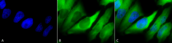
Immunocytochemistry/Immunofluorescence analysis using Mouse Anti-Hsp90 alpha/beta Monoclonal Antibody, Clone K41220A. Tissue: Cervical cancer cell line (HeLa). Species: Human. Fixation: 2% Formaldehyde for 20 min at RT. Primary Antibody: Mouse Anti-Hsp90 alpha/beta Monoclonal Antibody at 1:100 for 12 hours at 4°C. Secondary Antibody: FITC Goat Anti-Mouse (green) at 1:200 for 2 hours at RT. Counterstain: DAPI (blue) nuclear stain at 1:40000 for 2 hours at RT. Localization: Cytoplasm. Melanosome. Magnification: 100x. (A) DAPI (blue) nuclear stain. (B) Anti-Hsp90 alpha/beta Antibody. (C) Composite.
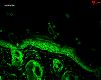
Immunohistochemistry analysis using Mouse Anti-Hsp90 alpha Monoclonal Antibody, Clone K41220A. Tissue: backskin. Species: Mouse. Fixation: Bouin's Fixative and paraffin-embedded. Primary Antibody: Mouse Anti-Hsp90 alpha Monoclonal Antibody at 1:100 for 1 hour at RT. Secondary Antibody: FITC Goat Anti-Mouse (green) at 1:50 for 1 hour at RT. Localization: Epidermis.
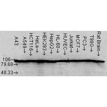
Western Blot analysis of Human Cell lysates showing detection of Hsp90 alpha protein using Mouse Anti-Hsp90 alpha Monoclonal Antibody, Clone K41220A. Load: 15 μg. Block: 1.5% BSA for 30 minutes at RT. Primary Antibody: Mouse Anti-Hsp90 alpha Monoclonal Antibody at 1:1000 for 2 hours at RT. Secondary Antibody: Sheep Anti-Mouse IgG: HRP for 1 hour at RT.
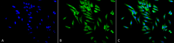
Immunocytochemistry/Immunofluorescence analysis using Mouse Anti-Hsp90 alpha/beta Monoclonal Antibody, Clone K41220A. Tissue: Cervical cancer cell line (HeLa). Species: Human. Fixation: 2% Formaldehyde for 20 min at RT. Primary Antibody: Mouse Anti-Hsp90 alpha/beta Monoclonal Antibody at 1:100 for 12 hours at 4°C. Secondary Antibody: FITC Goat Anti-Mouse (green) at 1:200 for 2 hours at RT. Counterstain: DAPI (blue) nuclear stain at 1:40000 for 2 hours at RT. Localization: Cytoplasm. Melanosome. Magnification: 20x. (A) DAPI (blue) nuclear stain. (B) Anti-Hsp90 alpha/beta Antibody. (C) Composite.
Quick Database Links
Gene Symbol
HSP90 alpha/beta
RefSeq (Protein):NP_001017963.2, NP_031381.2
UniProt Details
− No UniProt data available
NCBI Gene Details
− No NCBI Gene data available
NCBI Reference Sequences
−Associated Accession Numbers
Curated reference sequences for the gene transcript and protein product| Protein | NP_001017963.2, NP_031381.2 |
|---|
Documents Download
Datasheet
Product Information
Request a Document
Protocol Information
WB
Western Blot (IB, immunoblot)
IHC
Immunohistochemistry
IF
Immunofluorescence
ICC
Immunocytochemistry
ELISA
Enzyme-linked Immunosorbent Assay (EIA)
HSP90 alpha/beta Antibody (Biotin) (orb396201)

- 0.0
Based on 0 reviews
Participating in our Biorbyt product reviews program enables you to support fellow scientists by sharing your firsthand experience with our products.
Login to Submit a ReviewAvailable Sizes
Select a size below
Choose Conjugation or Carrier Free Version
Free Secondary Antibody (20 ul)0/0
Please add an antibody product to your cart first.