You have no items in your shopping cart.
Featured
Description
Research Area
Actina, Cancer Research, Cardiovascular Research, Signal Transduction, Tumor Biomarkers
Images & Validation
−Item 1 of 8
| Tested Applications | FC, IF, IHC-Fr, IHC-P, WB |
|---|---|
| Dilution Range | WB=1:500-2000, IHC-P=1:100-500, IHC-F=1:100-500, IF=1:100-500, Flow-Cyt=2μg/Test |
| Reactivity | Human, Rat |
| Predicted Reactivity | Bovine, Canine, Mouse, Porcine |
Related Conjugates & Formulations
−Key Properties
−| Antibody Type | Primary Antibody |
|---|---|
| Host | Rabbit |
| Clonality | Polyclonal |
| Isotype | IgG |
| Immunogen | KLH conjugated synthetic peptide derived from human HSP27 (101-205/205aa) |
| Target | HSPB1 |
| Molecular Weight | 27 kDa |
| Purification | Affinity purified by Protein A |
| Conjugation | Unconjugated |
Storage & Handling
−| Storage | Maintain refrigerated at 2-8°C for up to 2 weeks. For long term storage store at -20°C in small aliquots to prevent freeze-thaw cycles. |
|---|---|
| Form/Appearance | Liquid |
| Buffer/Preservatives | 0.01M TBS (pH7.4) with 1% rAlbumin, 0.02% Proclin300 and 50% Glycerol. |
| Concentration | 1mg/ml |
| Expiration Date | 12 months from date of receipt. |
| Disclaimer | For research use only |
Alternative Names
−CMT2F; HEL-S-102; HMN2B; HMND3; HS.76067; HSP27; HSP28; Hsp25; SRP27; 27kDa; HSPB1_CANLF; HSPB1; Heat shock 27 kDa protein (HSP 27); HSPB1_HUMAN; 28 kDa heat shock protein; Estrogen-regulated 24 kDa protein; Heat shock protein family B member 1; Stress-responsive protein 27 (SRP27); HSPB1_MOUSE; Growth-related 25 kDa protein; Heat shock 25 kDa protein (HSP 25); p25; HSPB1_RAT; heat shock protein family B (small) member 1; heat shock 27kD protein 1; heat shock 27kDa protein 1
Similar Products
−- Item 1 of 7
Hsp27/HSPB1 Rabbit Polyclonal Antibody [orb215991]
FC, ICC, IF, IHC, IHC-Fr, IP, WB
Human
Rabbit
Polyclonal
Unconjugated
100 μg - Item 1 of 7
HSP27 Polyclonal Antibody [orb1413400]
IF, IHC-P, WB
Human, Mouse, Rat
Rabbit
Polyclonal
Unconjugated
100 μl - Item 1 of 5
Phospho-HSP27 (Ser78) Rabbit Polyclonal Antibody [orb5481]
FC, ICC, IF, IHC-Fr, IHC-P, WB
Bovine, Canine, Equine, Rabbit
Human, Mouse, Rat
Rabbit
Polyclonal
Unconjugated
50 μl, 100 μl, 200 μl - Item 1 of 3
Phospho-HSP27 (Ser82) Rabbit Polyclonal Antibody [orb5482]
FC, IF, IHC-Fr, IHC-P
Bovine, Canine, Equine, Porcine, Rabbit
Human, Mouse, Rat
Rabbit
Polyclonal
Unconjugated
50 μl, 100 μl, 200 μl - Item 1 of 4
Phospho-HSP27 (Ser15) Rabbit Polyclonal Antibody [orb5483]
FC, ICC, IF, IHC-Fr, IHC-P, WB
Bovine, Sheep
Human
Rabbit
Polyclonal
Unconjugated
50 μl, 100 μl, 200 μl

Quality Guarantee
Explore bioreagents carefree to elevate your research. All our products are rigorously tested for performance. If a product does not perform as described on its datasheet, our scientific support team will provide expert troubleshooting, a prompt replacement, or a refund. For full details, please see our Terms & Conditions and Buying Guide. Contact us at [email protected].
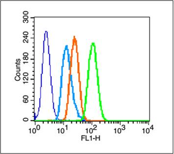
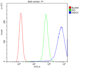
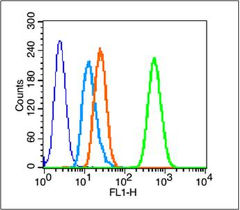
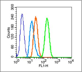
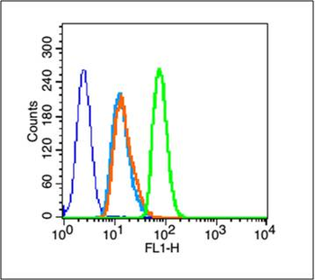
Blank control (blue line): A431 cells (blue). Primary Antibody (green line): Rabbit Anti-HSP27 antibody (orb10845), Dilution: 2 µg/10^6 cells, Isotype Control Antibody (orange line): Rabbit IgG. Secondary Antibody (white blue line): Goat anti-rabbit IgG-FITC, Dilution: 1 µg/Test. Protocol, The cells were fixed with 70% methanol (Overnight at 4°C) and then permeabilized with 90% ice-cold methanol for 20 min at -20°C. Cells stained with Primary Antibody for 30 min at room temperature. The cells were then incubated in 1 X PBS/2% BSA/10% goat serum to block non-specific protein-protein interactions followed by the antibody for 15 min at room temperature. The secondary antibody used for 40 min at room temperature. Acquisition of 20000 events was performed.
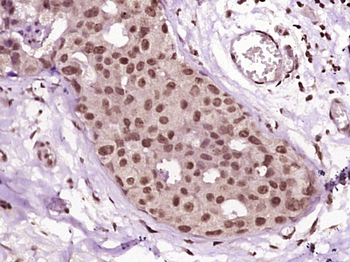
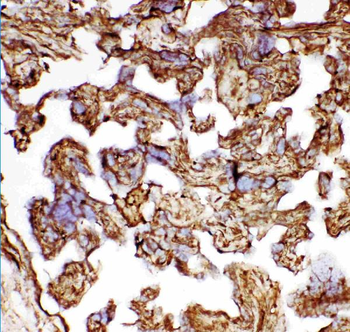
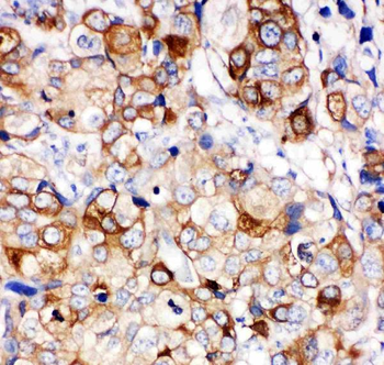
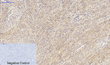
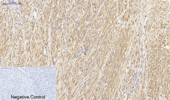
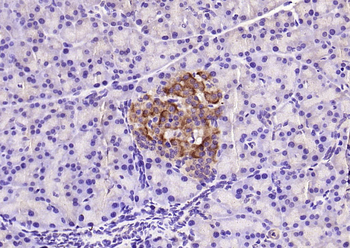
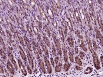
Paraformaldehyde-fixed, paraffin embedded (Human breast cancer), Antigen retrieval by boiling in sodium citrate buffer (pH6.0) for 15 min, Block endogenous peroxidase by 3% hydrogen peroxide for 20 minutes, Blocking buffer (normal goat serum) at 37°C for 30 min, Antibody incubation with (HSP27) Polyclonal Antibody, Unconjugated (orb10845) at 1:400 overnight at 4°C, followed by operating according to SP Kit (Rabbit) instructions and DAB staining.
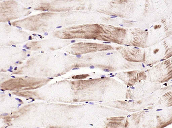
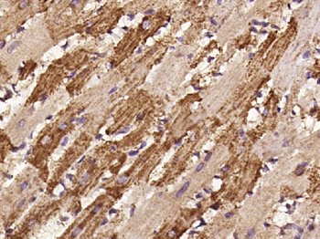
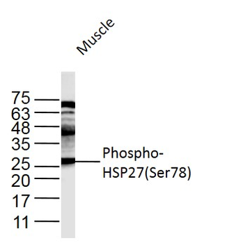
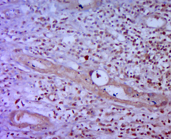
Paraformaldehyde-fixed, paraffin embedded (rat skeletal muscle), Antigen retrieval by boiling in sodium citrate buffer (pH6.0) for 15 min, Block endogenous peroxidase by 3% hydrogen peroxide for 20 minutes, Blocking buffer (normal goat serum) at 37°C for 30 min, Antibody incubation with (HSP27) Polyclonal Antibody, Unconjugated (orb10845) at 1:200 overnight at 4°C, followed by operating according to SP Kit (Rabbit) instructionsand DAB staining.
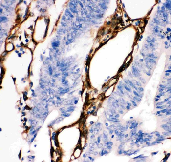
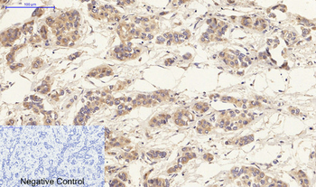
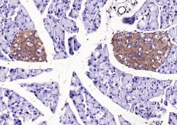
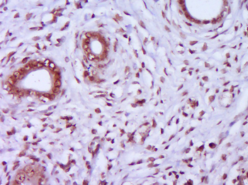
Paraformaldehyde-fixed, paraffin embedded (rat stomach tissue), Antigen retrieval by boiling in sodium citrate buffer (pH6.0) for 15 min, Block endogenous peroxidase by 3% hydrogen peroxide for 20 minutes, Blocking buffer (normal goat serum) at 37°C for 30 min, Antibody incubation with (HSP27) Polyclonal Antibody, Unconjugated (orb10845) at 1:400 overnight at 4°C, followed by operating according to SP Kit (Rabbit) instructionsand DAB staining.
Paraformaldehyde-fixed, paraffin embedded (Rat uterus), Antigen retrieval by boiling in sodium citrate buffer (pH6.0) for 15 min, Block endogenous peroxidase by 3% hydrogen peroxide for 20 minutes, Blocking buffer (normal goat serum) at 37°C for 30 min, Antibody incubation with (HSP27) Polyclonal Antibody, Unconjugated (orb10845) at 1:400 overnight at 4°C, followed by operating according to SP Kit (Rabbit) instructionsand DAB staining.
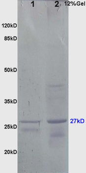
Sample: Brain (Mouse) lysate at 30 ug, Liver (Mouse) lysate at 30 ug, Primary: Anti-HSP-27 (orb10845) at 1:200, Secondary: AP conjugated Goat Anti-Rabbit IgG at 1:3000 dilution, NBT/BCIP staining, Predicted band size: 27kD, Observed band size: 27kD.
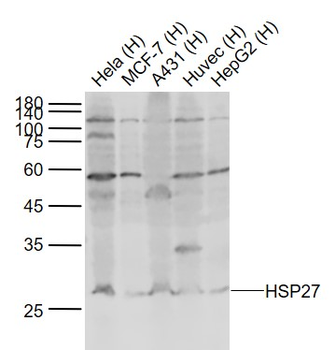
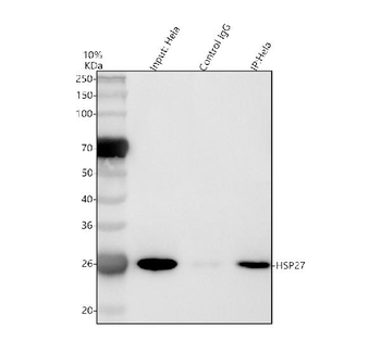
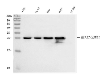
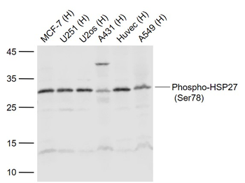
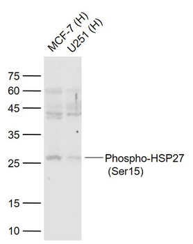
Sample: Lane 1: Hela (Human) Cell Lysate at 30 ug, Lane 2: MCF-7 (Human) Cell Lysate at 30 ug, Lane 3: A431 (Human) Cell Lysate at 30 ug, Lane 4: Huvec (Human) Cell Lysate at 30 ug, Lane 5: HepG2 (Human) Cell Lysate at 30 ug, Primary: Anti-HSP27 (orb10845) at 1/1000 dilution, Secondary: IRDye800CW Goat Anti-Rabbit IgG at 1/20000 dilution, Predicted band size: 27-30 kD, Observed band size: 27 kD.
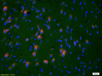
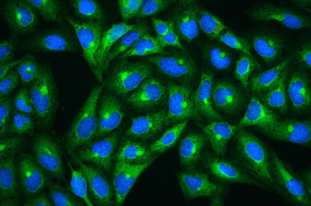
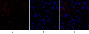
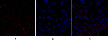
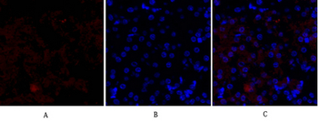
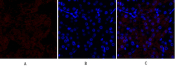
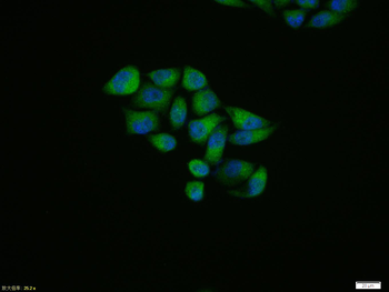
Tissue/Cell: rat brain tissue, 4% Paraformaldehyde-fixed and paraffin-embedded, Antigen retrieval: citrate buffer (0.01M, pH6.0), Boiling bathing for 15 min, Blocking buffer (normal goat serum) at 37°C for 20 min, Incubation: Anti-HSP-27 Polyclonal Antibody, Unconjugated (orb10845) 1:200, overnight at 4°C, The secondary antibody was Goat Anti-Rabbit IgG, Cy3 conjugated (orb868589) used at 1:200 dilution for 40 minutes at 37°C. DAPI (5 ug/ml, blue) was used to stain the cell nuclei.
Quick Database Links
Gene Symbol
HSPB1
UniProt
UniProt Details
− No UniProt data available
Documents Download
Datasheet
Product Information
Request a Document
Protocol Information
WB
Western Blot (IB, immunoblot)
IHC-P
Immunohistochemistry Paraffin
IHC-Fr
Immunohistochemistry Frozen
FC
Flow Cytometry
IF
Immunofluorescence
HSP27 Rabbit Polyclonal Antibody (orb10845)

- 0.0
Based on 0 reviews
Participating in our Biorbyt product reviews program enables you to support fellow scientists by sharing your firsthand experience with our products.
Login to Submit a ReviewAvailable Sizes
Select a size below
Free Secondary Antibody (20 ul)0/0
Please add an antibody product to your cart first.