You have no items in your shopping cart.
Featured
Description
Research Area
Cancer Research, Cell Biology, Epidermal Growth Factor, Receptor Tyrosne Kinases, Tumor Biology
Images & Validation
−Item 1 of 5
| Tested Applications | ICC, IF, IHC-Fr, IHC-P, WB |
|---|---|
| Dilution Range | WB=1:500-2000, IHC-P=1:100-500, IHC-F=1:100-500, ICC/IF=1:100-500, IF=1:100-500 |
| Reactivity | Human |
| Predicted Reactivity | Canine, Equine |
Related Conjugates & Formulations
−Key Properties
−| Antibody Type | Primary Antibody |
|---|---|
| Host | Mouse |
| Clonality | Monoclonal |
| Isotype | IgG |
| Clone No. | 1A4 |
| Immunogen | KLH conjugated synthetic peptide derived from human HER2 receptor (151-250/1255aa) |
| Target | ERBB2 |
| Molecular Weight | 138 kDa |
| Purification | Affinity purified by Protein A |
| Conjugation | Unconjugated |
Storage & Handling
−| Storage | Maintain refrigerated at 2-8°C for up to 2 weeks. For long term storage store at -20°C in small aliquots to prevent freeze-thaw cycles. |
|---|---|
| Form/Appearance | Liquid |
| Buffer/Preservatives | 0.01M TBS (pH7.4) with 1% rAlbumin, 0.02% Proclin300 and 50% Glycerol. |
| Concentration | 1mg/ml |
| Expiration Date | 12 months from date of receipt. |
| Disclaimer | For research use only |
Alternative Names
−erbB-2 isoform 2; HER2 receptor; Erbb2 protein; CerbB2; c erb B2; C erbB 2; C-erbB2; CD340; Erb B2; erbb2; HER 2; HER 2/neu; Her2/neu; Herstatin; MLN 19; MLN19; NEU; NEU Proto Oncogene; Neuro Glioblastoma Derived Oncogene Homolog; NGL; p185 ErbB2; p185erbB2; Receptor Protein Tyrosine Kinase ErbB2 Precursor; Receptor tyrosine protein kinase erbB 2; TKR1; Tyrosine kinase type cell surface receptor HER2; v erb b2 erythroblastic leukemia viral oncogene homolog 2 neuro/glioblastoma derived oncogene homolog(avian); ERBB2_HUMAN; Receptor tyrosine-protein kinase erbB-2; Metastatic lymph node gene 19 protein; Proto-oncogene Neu; Proto-oncogene c-ErbB-2.
Similar Products
−- Item 1 of 5
C-erbB-2/HER2 Mouse Monoclonal Antibody [orb499961]
IF, IHC-Fr, IHC-P, WB
Mouse, Rat
Human
Mouse
Monoclonal
Unconjugated
50 μl, 100 μl, 200 μl, 200 μg - Item 1 of 3
- Item 1 of 2
- Item 1 of 2
- Item 1 of 2
![Anti-HER2/neu [SER4]](/images/pub/media/catalog/product/NewWebsite/35/orb669729_1.png)
![Anti-HER2/neu [SER4]](/images/pub/media/catalog/product/NewWebsite/35/orb669729_2.png)
![Anti-HER2/neu [SER4]](/images/pub/media/catalog/product/NewWebsite/35/orb669729_3.png)


![Anti-c-erbB-2 [FWP51]](/images/pub/media/catalog/product/NewWebsite/35/orb669758_1.png)
![Anti-c-erbB-2 [FWP51]](/images/pub/media/catalog/product/NewWebsite/35/orb669758_2.png)
![Anti-c-erbB-2 [FWP51]](/images/pub/media/catalog/product/NewWebsite/35/orb669759_1.png)
![Anti-c-erbB-2 [FWP51]](/images/pub/media/catalog/product/NewWebsite/35/orb669759_2.png)
Quality Guarantee
Explore bioreagents carefree to elevate your research. All our products are rigorously tested for performance. If a product does not perform as described on its datasheet, our scientific support team will provide expert troubleshooting, a prompt replacement, or a refund. For full details, please see our Terms & Conditions and Buying Guide. Contact us at [email protected].
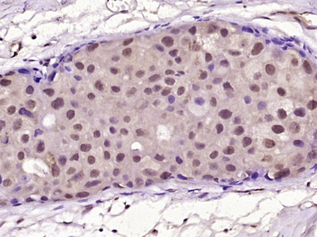
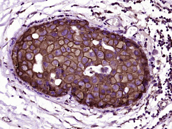
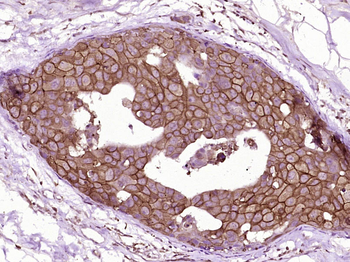
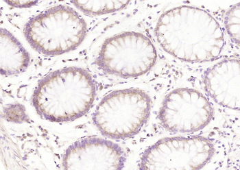
Paraformaldehyde-fixed, paraffin embedded (Human breast carcinoma), Antigen retrieval by boiling in sodium citrate buffer (pH6.0) for 15 min, Block endogenous peroxidase by 3% hydrogen peroxide for 20 minutes, Blocking buffer (normal goat serum) at 37°C for 30 min, Antibody incubation with (HER2 receptor) Monoclonal Antibody, Unconjugated at 1:400 overnight at 4°C, followed by a conjugated secondary for 20 minutes and DAB staining.
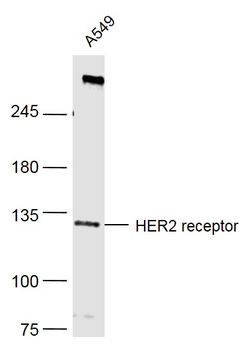
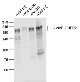
Sample: A549 (Human) Cell Lysate at 40 ug, Primary: Anti-HER2 receptor at 1/500 dilution, Secondary: IRDye800CW Goat Anti-Mouse IgG at 1/20000 dilution, Predicted band size: 138 kD, Observed band size: 134 kD.
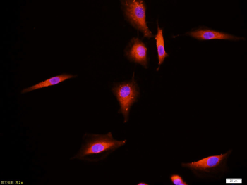
Tissue/cell:A549 cell, 4% Paraformaldehyde-fixed, Triton X-100 at room temperature for 20 min, Blocking buffer (normal goat serum) at 37°C for 20 min, Antibody incubation with (HER2 receptor) monoclonal Antibody, Unconjugated (orb101953) 1:100, 90 minutes at 37°C, followed by a CY3 conjugated Goat Anti-Mouse IgG antibody at 37°C for 90 minutes, DAPI (blue) was used to stain the cell nuclei.
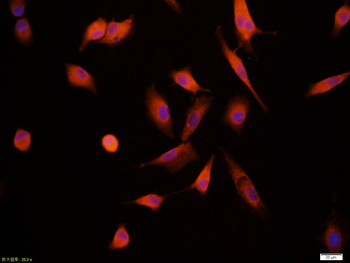
Tissue/cell:A549 cell, 4% Paraformaldehyde-fixed, Triton X-100 at room temperature for 20 min, Blocking buffer (normal goat serum) at 37°C for 20 min, Antibody incubation with (HER2 receptor) monoclonal Antibody, Unconjugated (orb101953) 1:100, 90 minutes at 37°C, followed by a CY3 conjugated Goat Anti-Mouse IgG antibody at 37°C for 90 minutes, DAPI (blue) was used to stain the cell nuclei.
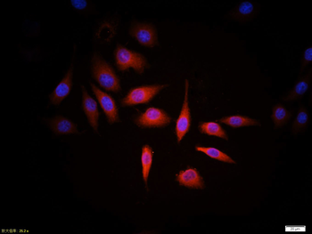
Tissue/cell:MCF7 cell, 4% Paraformaldehyde-fixed, Triton X-100 at room temperature for 20 min, Blocking buffer (normal goat serum) at 37°C for 20 min, Antibody incubation with (HER2 receptor) monoclonal Antibody, Unconjugated (orb101953) 1:100, 90 minutes at 37°C, followed by a CY3 conjugated Goat Anti-Mouse IgG antibody at 37°C for 90 minutes, DAPI (blue) was used to stain the cell nuclei.
Quick Database Links
Gene Symbol
ERBB2
UniProt
UniProt Details
− No UniProt data available
Documents Download
Datasheet
Product Information
Request a Document
Protocol Information
WB
Western Blot (IB, immunoblot)
IHC-P
Immunohistochemistry Paraffin
IHC-Fr
Immunohistochemistry Frozen
IF
Immunofluorescence
ICC
Immunocytochemistry
Caceres, S. et al. Canine cell line, IPC-366, as a good model for the study of inflammatory breast cancer Vet Comp Oncol, 15, 980-995 (2017)
HER2 Mouse Monoclonal Antibody (orb101953)

- 0.0
Based on 0 reviews
Participating in our Biorbyt product reviews program enables you to support fellow scientists by sharing your firsthand experience with our products.
Login to Submit a Review