You have no items in your shopping cart.
Description
Images & Validation
−Item 1 of 6
| Tested Applications | ELISA, IF, IHC, Multiplex Assay, WB |
|---|---|
| Dilution Range | ELISA: 1:10,000 - 1:50,000, IHC: 1:200 - 1:1,000, IF: 10ug / ml, WB: 1:1,000 - 1:5,000 |
| Reactivity | Human |
| Application Notes |
Key Properties
−| Antibody Type | Primary Antibody |
|---|---|
| Host | Rabbit |
| Clonality | Polyclonal |
| Isotype | IgG |
| Immunogen | Anti-HDAC-1 antibody was prepared from whole rabbit serum produced by repeated immunizations with a synthetic peptide corresponding to a C-Terminal region near amino acids 450-482 of Human HDAC-1. |
| Target | HDAC1 |
| Purity | Anti-HDAC-1 antibody is directed against human HDAC-1 protein. HDAC-1 antibody was affinity purified from monospecific antiserum by immunoaffinity purification. A BLAST analysis was used to suggest reactivity with this protein from human, mouse, rat and chimpanzee sources based on 100% homology for the immunogen sequence. Cross reactivity may occur with HDAC-1 from bovine (82% homology) and chicken (80% homology) sources. Cross reactivity with HDAC-1 homologues from other sources has not been determined. |
| Conjugation | Unconjugated |
Storage & Handling
−| Storage | Store vial at -20° C or below prior to opening. This vial contains a relatively low volume of reagent (25 µL). To minimize loss of volume dilute 1:10 by adding 225 µL of the buffer stated above directly to the vial. Recap, mix thoroughly and briefly centrifuge to collect the volume at the bottom of the vial. Use this intermediate dilution when calculating final dilutions as recommended below. Store the vial at -20°C or below after dilution. Avoid cycles of freezing and thawing. |
|---|---|
| Form/Appearance | Liquid (sterile filtered) |
| Buffer/Preservatives | Preservative: 0.1% (w/v) Sodium Azide. Stabilizer: None; Buffer: 0.02 M Potassium Phosphate, 0.15 M Sodium Chloride, pH 7.2 |
| Concentration | 1.0 mg/mL |
| Expiration Date | 12 months from date of receipt. |
| Dry Ice Shipping | Please note: This product requires shipment on dry ice. A dry ice surcharge will apply. |
| Disclaimer | For research use only |
Alternative Names
−rabbit anti-HDAC-1 antibody, HDAC1, HDAC 1, HD 1 antibody, histone deacetylase 1 antibody, Histone deacetylase-1, HD1 antibody, RPD3L1, reduced potassium dependency yeast homolog like 1 antibody
Similar Products
−- Item 1 of 4
HDAC1 Rabbit Polyclonal Antibody [orb10812]
FC, IF, IHC-Fr, IHC-P, WB
Bovine, Canine, Guinea pig, Porcine, Rat, Sheep
Human, Mouse
Rabbit
Polyclonal
Unconjugated
50 μl, 100 μl, 200 μl - Item 1 of 6
HDAC1 Rabbit Polyclonal Antibody [orb574159]
IHC, WB
Bovine, Canine, Equine, Guinea pig, Mouse, Rabbit, Rat, Yeast, Zebrafish
Human
Rabbit
Polyclonal
Unconjugated
100 μl - Item 1 of 4
HDAC1 Antibody [orb1410123]
ELISA, FC, IF
Human
Mouse
Monoclonal
Unconjugated
100 μg (without BSA and Azide), 20 μg, 100 μg - Item 1 of 6
- Item 1 of 6
HDAC1 Antibody [orb345508]
ELISA, IF, IHC, Multiplex Assay, WB
Human
Rabbit
Polyclonal
Unconjugated
100 μg


Quality Guarantee
Explore bioreagents carefree to elevate your research. All our products are rigorously tested for performance. If a product does not perform as described on its datasheet, our scientific support team will provide expert troubleshooting, a prompt replacement, or a refund. For full details, please see our Terms & Conditions and Buying Guide. Contact us at [email protected].
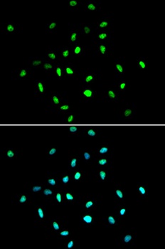
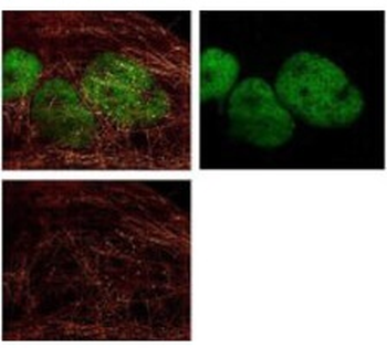
Immunofluorescence Microscopy of Rabbit Anti-HDAC-1 antibody. Fixation: 0.5% PFA. Antigen retrieval: not required. Primary antibody: HDAC-1 antibody at 10 µg/ml for 1 h at RT. Secondary antibody: rabbit secondary antibody at 1:10000 for 45 min at RT. Localization: HDAC-1 is nuclear. Staining: HDAC-1 was used with Atto 425 (shown in red). Anti-Keratin monoclonal antibody was used with Dylight 488 (shown in green) to detect Keratin.
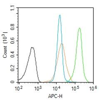
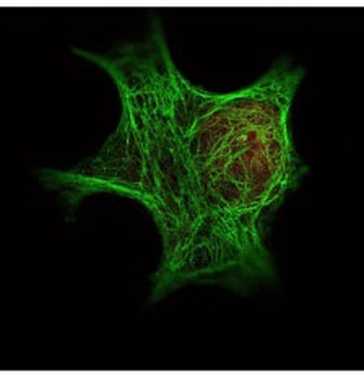
Immunofluorescence Microscopy of Rabbit anti-HDAC1 Antibody. Tissue: A431 cells. Fixation: methanol. Antigen retrieval: blocked with normal goat serum. Primary antibody: HDAC1 antibody at 4 µg/ml for 1 h at RT. Secondary antibody: rabbit secondary antibody at 0.2 µg/ml for 45 min at RT. Localization: HDAC1 is nuclear. Staining: HDAC1 as green fluorescent signal. A-tubulin monoclonal antibody detected with ATTO 425 (colored RED). 2-color STED image, Leica Microsystems.

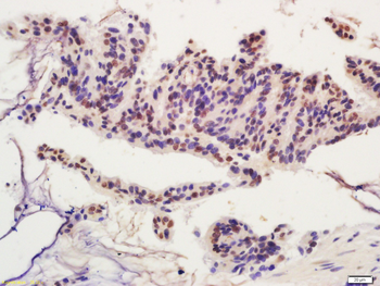
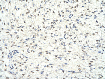
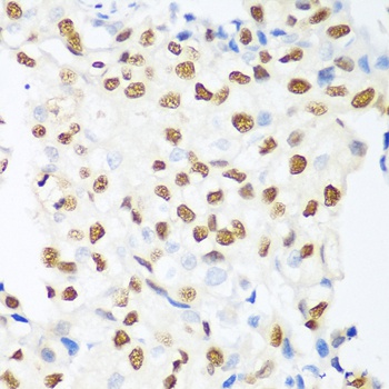
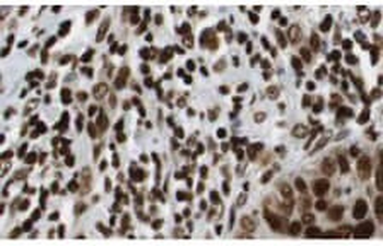
Immunohistochemistry of Rabbit Anti-HDAC-1 Antibody. Tissue: human lung tissue. Fixation: formalin fixed paraffin embedded. Antigen retrieval: not required. Primary antibody: HDAC-1 antibody at 10 µg/ml for 1 h at RT. Secondary antibody: Peroxidase rabbit secondary antibody at 1:10000 for 45 min at RT. Localization: HDAC-1 is nuclear. Staining: HDAC-1 as brown color indicates presence of protein, blue color shows cell nuclei.

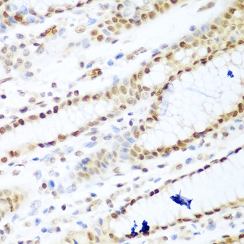
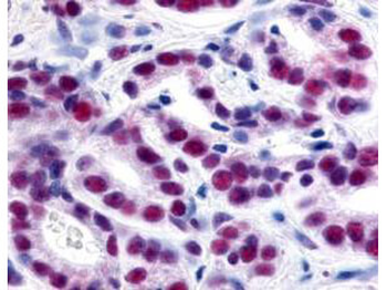
Immunohistochemistry of Rabbit Anti-HDAC-1 Antibody. Tissue: human prostate cancer tissue. Fixation: formalin fixed paraffin embedded. Antigen retrieval: not required. Primary antibody: HDAC-1 antibody at 1:500 for 1 h at RT. Secondary antibody: Peroxidase rabbit secondary antibody at 1:10000 for 45 min at RT. Localization: HDAC-1 is nuclear. Staining: HDAC-1 precipitated purple with blue counterstain.
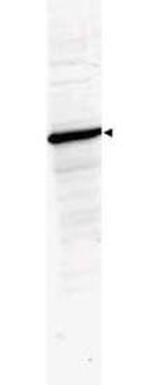
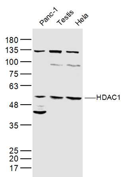
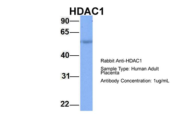
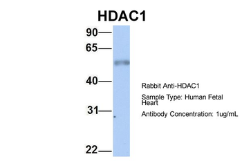
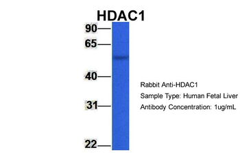
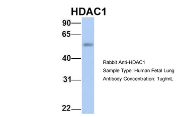
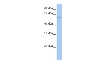
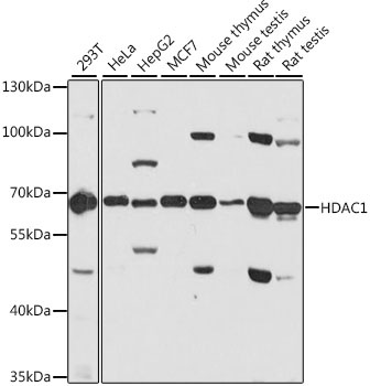
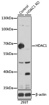
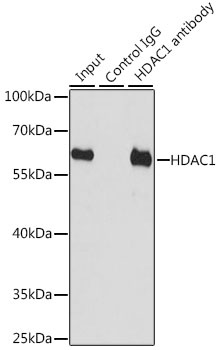
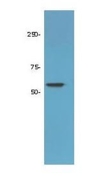
Western Blot of Rabbit Anti-HDAC-1 Antibody. Lane 1: 293 whole cell lysate (p/n orb348669). Load: 35 µg per lane. Primary antibody: HDAC-1 antibody at 1:3500 for overnight at 4°C. Secondary antibody: IRDye800™ rabbit secondary antibody at 1:10000 for 45 min at RT. Block: 5% BLOTTO (p/n orb348624) overnight at 4°C. Predicted/Observed size: ~65 kDa corresponding to human HDAC1. Other band(s): none.

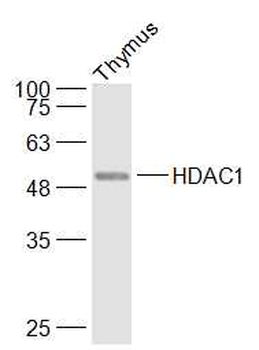
Western Blot of Rabbit Anti-HDAC-1 Antibody. Lane 1: LNCaP prostate cancer cells. Load: 50 µg per lane. Primary antibody: HDAC-1 antibody at 1:1000 for overnight at 4°C. Secondary antibody: IRDye800™ rabbit secondary antibody at 1:10000 for 45 min at RT. Block: 5% BLOTTO overnight at 4°C. Predicted/Observed size: 55kDa for HDAC-1.
Documents Download
Datasheet
Product Information
Request a Document
Protocol Information
WB
Western Blot (IB, immunoblot)
IHC
Immunohistochemistry
IF
Immunofluorescence
ELISA
Enzyme-linked Immunosorbent Assay (EIA)
HDAC1 Antibody (orb345509)

- 0.0
Based on 0 reviews
Participating in our Biorbyt product reviews program enables you to support fellow scientists by sharing your firsthand experience with our products.
Login to Submit a ReviewAvailable Sizes
Select a size below
Free Secondary Antibody (20 ul)0/0
Please add an antibody product to your cart first.