You have no items in your shopping cart.
Description
Images & Validation
−Item 1 of 6
| Tested Applications | ELISA, IF, IHC, Multiplex Assay, WB |
|---|---|
| Dilution Range | ELISA: 1:10,000 - 1:50,000, IHC: 1:200 - 1:1,000, IF: 10ug / ml, WB: 1:1,000 - 1:5,000 |
| Reactivity | Human |
| Application Notes |
Key Properties
−| Antibody Type | Primary Antibody |
|---|---|
| Host | Rabbit |
| Clonality | Polyclonal |
| Isotype | IgG |
| Immunogen | Anti-HDAC-1 antibody was prepared from whole rabbit serum produced by repeated immunizations with a synthetic peptide corresponding to a C-Terminal region near amino acids 450-482 of Human HDAC-1. |
| Target | HDAC1 |
| Purity | Anti-HDAC-1 antibody is directed against human HDAC-1 protein. HDAC-1 antibody was affinity purified from monospecific antiserum by immunoaffinity purification. A BLAST analysis was used to suggest reactivity with this protein from human, mouse, rat and chimpanzee sources based on 100% homology for the immunogen sequence. Cross reactivity may occur with HDAC-1 from bovine (82% homology) and chicken (80% homology) sources. Cross reactivity with HDAC-1 homologues from other sources has not been determined. |
| Conjugation | Unconjugated |
Storage & Handling
−| Storage | Store vial at -20° C prior to opening. Aliquot contents and freeze at -20° C or below for extended storage. Avoid cycles of freezing and thawing. Centrifuge product if not completely clear after standing at room temperature. This product is stable for several weeks at 4° C as an undiluted liquid. Dilute only prior to immediate use. |
|---|---|
| Form/Appearance | Liquid (sterile filtered) |
| Buffer/Preservatives | Preservative: 0.01% (w/v) Sodium Azide. Stabilizer: None; Buffer: 0.02 M Potassium Phosphate, 0.15 M Sodium Chloride, pH 7.2 |
| Concentration | 1.33 mg/mL |
| Expiration Date | 12 months from date of receipt. |
| Dry Ice Shipping | Please note: This product requires shipment on dry ice. A dry ice surcharge will apply. |
| Disclaimer | For research use only |
Alternative Names
−rabbit anti-HDAC-1 antibody, HDAC1, HDAC 1, HD 1 antibody, histone deacetylase 1 antibody, Histone deacetylase-1, HD1 antibody, RPD3L1, reduced potassium dependency yeast homolog like 1 antibody
Similar Products
−- Item 1 of 4
HDAC1 Rabbit Polyclonal Antibody [orb10812]
FC, IF, IHC-Fr, IHC-P, WB
Bovine, Canine, Guinea pig, Porcine, Rat, Sheep
Human, Mouse
Rabbit
Polyclonal
Unconjugated
50 μl, 100 μl, 200 μl - Item 1 of 6
HDAC1 Rabbit Polyclonal Antibody [orb574159]
IHC, WB
Bovine, Canine, Equine, Guinea pig, Mouse, Rabbit, Rat, Yeast, Zebrafish
Human
Rabbit
Polyclonal
Unconjugated
100 μl - Item 1 of 4
HDAC1 Antibody [orb1410123]
ELISA, FC, IF
Human
Mouse
Monoclonal
Unconjugated
100 μg (without BSA and Azide), 20 μg, 100 μg - Item 1 of 6
- Item 1 of 6
HDAC1 Antibody [orb345509]
ELISA, IF, IHC, Multiplex Assay, WB
Human
Rabbit
Polyclonal
Unconjugated
25 μl


Quality Guarantee
Explore bioreagents carefree to elevate your research. All our products are rigorously tested for performance. If a product does not perform as described on its datasheet, our scientific support team will provide expert troubleshooting, a prompt replacement, or a refund. For full details, please see our Terms & Conditions and Buying Guide. Contact us at [email protected].

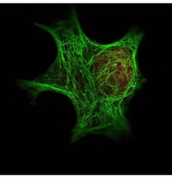
Immunofluorescence Microscopy of Rabbit Anti-HDAC-1 antibody. Fixation: 0.5% PFA. Antigen retrieval: not required. Primary antibody: HDAC-1 antibody at 10 µg/ml for 1 h at RT. Secondary antibody: rabbit secondary antibody at 1:10000 for 45 min at RT. Localization: HDAC-1 is nuclear. Staining: HDAC-1 was used with Atto 425 (shown in red). Anti-Keratin monoclonal antibody was used with Dylight 488 (shown in green) to detect Keratin.

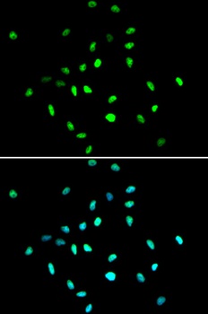
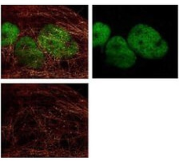
Immunofluorescence Microscopy of Rabbit anti-HDAC1 Antibody. Tissue: A431 cells. Fixation: methanol. Antigen retrieval: blocked with normal goat serum. Primary antibody: HDAC1 antibody at 4 µg/ml for 1 h at RT. Secondary antibody: rabbit secondary antibody at 0.2 µg/ml for 45 min at RT. Localization: HDAC1 is nuclear. Staining: HDAC1 as green fluorescent signal. A-tubulin monoclonal antibody detected with ATTO 425 (colored RED). 2-color STED image, Leica Microsystems.
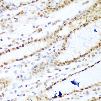
Immunohistochemistry of Rabbit Anti-HDAC-1 Antibody. Tissue: human lung tissue. Fixation: formalin fixed paraffin embedded. Antigen retrieval: not required. Primary antibody: HDAC-1 antibody at 10 µg/ml for 1 h at RT. Secondary antibody: Peroxidase rabbit secondary antibody at 1:10000 for 45 min at RT. Localization: HDAC-1 is nuclear. Staining: HDAC-1 as brown color indicates presence of protein, blue color shows cell nuclei.
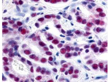
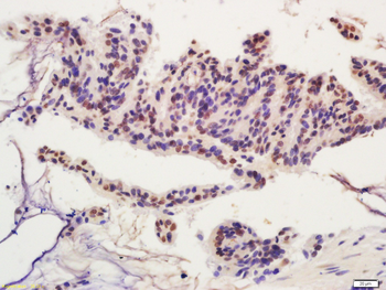
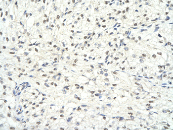
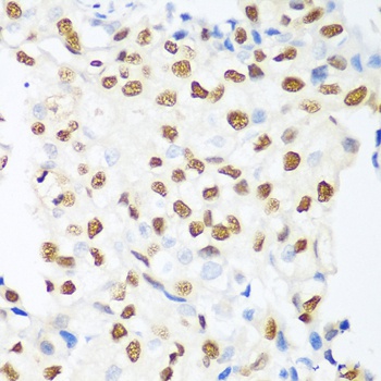
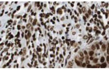
Immunohistochemistry of Rabbit Anti-HDAC-1 Antibody. Tissue: human prostate cancer tissue. Fixation: formalin fixed paraffin embedded. Antigen retrieval: not required. Primary antibody: HDAC-1 antibody at 1:500 for 1 h at RT. Secondary antibody: Peroxidase rabbit secondary antibody at 1:10000 for 45 min at RT. Localization: HDAC-1 is nuclear. Staining: HDAC-1 precipitated purple with blue counterstain.

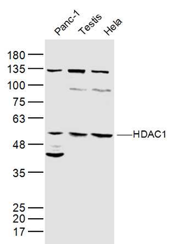
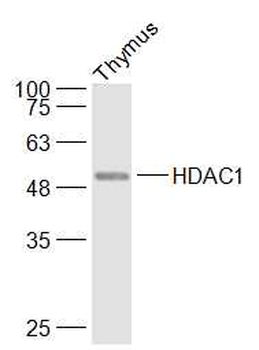
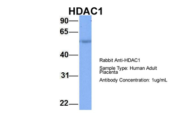
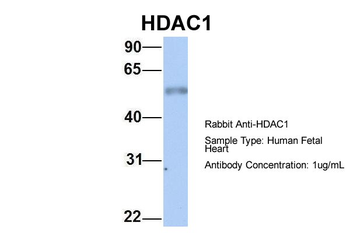
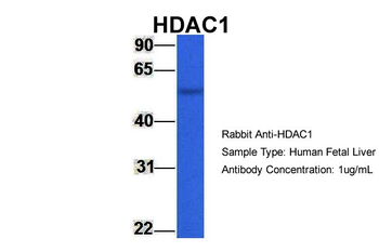
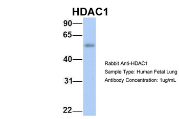
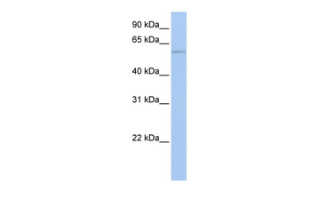
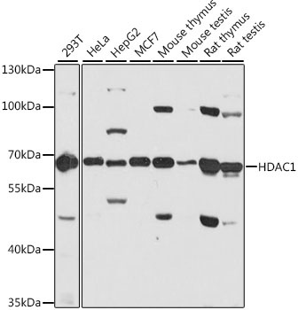
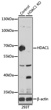
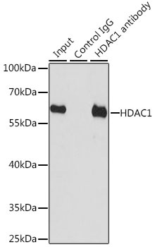
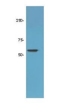
Western Blot of Rabbit Anti-HDAC-1 Antibody. Lane 1: 293 whole cell lysate (p/n orb348669). Load: 35 µg per lane. Primary antibody: HDAC-1 antibody at 1:3500 for overnight at 4°C. Secondary antibody: IRDye800™ rabbit secondary antibody at 1:10000 for 45 min at RT. Block: 5% BLOTTO (p/n orb348624) overnight at 4°C. Predicted/Observed size: ~65 kDa corresponding to human HDAC1. Other band(s): none.
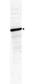
Western Blot of Rabbit Anti-HDAC-1 Antibody. Lane 1: LNCaP prostate cancer cells. Load: 50 µg per lane. Primary antibody: HDAC-1 antibody at 1:1000 for overnight at 4°C. Secondary antibody: IRDye800™ rabbit secondary antibody at 1:10000 for 45 min at RT. Block: 5% BLOTTO overnight at 4°C. Predicted/Observed size: 55kDa for HDAC-1.
Documents Download
Datasheet
Product Information
Request a Document
Protocol Information
WB
Western Blot (IB, immunoblot)
IHC
Immunohistochemistry
IF
Immunofluorescence
ELISA
Enzyme-linked Immunosorbent Assay (EIA)
HDAC1 Antibody (orb345508)

- 0.0
Based on 0 reviews
Participating in our Biorbyt product reviews program enables you to support fellow scientists by sharing your firsthand experience with our products.
Login to Submit a ReviewAvailable Sizes
Select a size below
Choose Conjugation or Carrier Free Version
Free Secondary Antibody (20 ul)0/0
Please add an antibody product to your cart first.