You have no items in your shopping cart.
KO/KD
Validated
Validated
Description
Research Area
Cardiovascular Research, Cell Biology, Infectious Disease & Virology, Signal Transduction
Images & Validation
−Item 1 of 4
| Tested Applications | IF, KO/KD Validated, WB |
|---|---|
| Dilution Range | IF - 1:10-50, WB - 1:2000 |
| Reactivity | Human, Mouse |
Key Properties
−| Host | Rabbit |
|---|---|
| Clonality | Polyclonal |
| Isotype | Rabbit IgG |
| Immunogen | This HDAC1 antibody is generated from rabbits immunized with a KLH conjugated synthetic peptide between 449-482 amino acids from the C-terminal region of human HDAC1. Antigen Region: 449-482 aa. |
| Target | HDAC1 {ECO:0000303|PubMed:10846170, ECO:0000312|HGNC:HGNC:4852} |
| Molecular Weight | 55103 Da |
| Conjugation | Unconjugated |
Storage & Handling
−| Storage | Maintain refrigerated at 2-8°C for up to 2 weeks. For long term storage store at -20°C in small aliquots to prevent freeze-thaw cycles |
|---|---|
| Form/Appearance | Purified polyclonal antibody supplied in PBS with 0.09% (W/V) sodium azide. This antibody is prepared by Saturated Ammonium Sulfate (SAS) precipitation followed by dialysis against PBS. |
| Expiration Date | 12 months from date of receipt. |
| Disclaimer | For research use only |
Alternative Names
−Histone deacetylase 1, HD1, HDAC1, RPD3L1
Similar Products
−
- Item 1 of 1
HDAC1 Antibody(C-term) [orb1788387]
WB
Mouse
Human, Rat
Rabbit
Polyclonal
Unconjugated
Quality Guarantee
Explore bioreagents carefree to elevate your research. All our products are rigorously tested for performance. If a product does not perform as described on its datasheet, our scientific support team will provide expert troubleshooting, a prompt replacement, or a refund. For full details, please see our Terms & Conditions and Buying Guide. Contact us at [email protected].
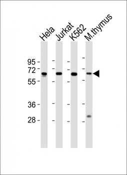
All lanes: Anti-HDAC1 Antibody (K464) at 1:2000 dilution. Lane 1: Hela whole cell lysates. Lane 2: Jurkat whole cell lysates. Lane 3: K562 whole cell lysates. Lane 4: mouse thymus lysates.Lysates/proteins at 20 µg per lane. Secondary Goat Anti-Rabbit IgG, (H+L), Peroxidase conjugated at 1/10000 dilution. Predicted band size: 55 kDa. Blocking/Dilution buffer: 5% NFDM/TBST.

Fluorescent image of Hela cell stained with HDAC1 Antibody (C-term). Hela cells were fixed with 4% PFA (20 min), permeabilized with Triton X-100 (0.1%, 10 min), then incubated with HDAC1 primary antibody (1:25, 1 h at 37°C). For secondary antibody, Alexa Fluor 488 conjugated donkey anti-rabbit antibody (green) was used (1:400, 50 min at 37°C). Cytoplasmic actin was counterstained with Alexa Fluor 555 (red) conjugated Phalloidin (7 units/ml, 1 h at 37°C). HDAC1 immunoreactivity is localized to Nucleus significantly.
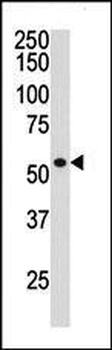
The anti-HDAC1 Pab is used in Western blot to detect HDAC1 in ZR-75-1 cell lysate.
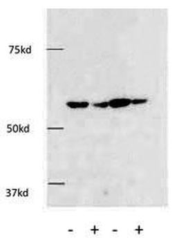
The anti-HDAC1 Pab was used in Western Blot to detect HDAC1 in HEK293 cells.Knockdown of HDAC1 using siRNA against HDAC1 showed a significant decrease of HDAC1 protein using this anti-HDAC1 Pab in HEK293 cells.
Quick Database Links
Gene Symbol
HDAC1 {ECO:0000303|PubMed:10846170, ECO:0000312|HGNC:HGNC:4852}
UniProt
RefSeq (Protein):NP_004955.2
UniProt Details
− No UniProt data available
NCBI Reference Sequences
−Associated Accession Numbers
Curated reference sequences for the gene transcript and protein product| Protein | NP_004955.2 |
|---|
Documents Download
Datasheet
Product Information
Request a Document
Protocol Information
WB
Western Blot (IB, immunoblot)
IF
Immunofluorescence
HDAC1 Antibody (C-term) (orb1938525)

- 0.0
Based on 0 reviews
Participating in our Biorbyt product reviews program enables you to support fellow scientists by sharing your firsthand experience with our products.
Login to Submit a ReviewAvailable Sizes
Select a size below
Choose Conjugation or Carrier Free Version
Free Secondary Antibody (20 ul)0/0
Please add an antibody product to your cart first.