You have no items in your shopping cart.
Featured
Description
Research Area
Cardiovascular Research, Neuroscience, Pharmacology & Drug Discovery
Images & Validation
−Item 1 of 6
| Tested Applications | AM, ICC, IF, IHC, IP, WB |
|---|---|
| Dilution Range | WB (1:1000), IHC-P (1:1000), ICC/IF (1:100) |
| Reactivity | Mouse, Rat |
| Application Notes |
Key Properties
−| Antibody Type | Recombinant Antibody |
|---|---|
| Host | Mouse |
| Clonality | Recombinant |
| Isotype | IgG1 |
| Clone No. | S71 |
| Immunogen | Fusion protein amino acids 761-863 (cytoplasmic C-terminus) of rat HCN2 |
| Target | HCN2 |
| Molecular Weight | 95kDa |
| Purification | Protein G Purified |
| Conjugation | Unconjugated |
Storage & Handling
−| Storage | Maintain refrigerated at 2-8°C for up to 2 weeks. For long term storage store at -20°C in small aliquots to prevent freeze-thaw cycles. |
|---|---|
| Buffer/Preservatives | PBS pH 7.4, 50% glycerol, 0.09% sodium azide. Storage buffer changes when conjugated. |
| Concentration | 1 mg/ml |
| Expiration Date | 12 months from date of receipt. |
| Disclaimer | For research use only |
Alternative Names
−BCNG2, HAC1, brain cyclic nucleotide gated channel 2, Potassium/sodium hyperpolarization-activated cyclic nucleotide-gated channel
Similar Products
−- Item 1 of 10
Hcn2 Rabbit Polyclonal Antibody [orb1819380]
ELISA, FC, IHC, WB
Mouse, Rat
Rabbit
Polyclonal
Unconjugated
100 μg - Item 1 of 6
- Item 1 of 6
- Item 1 of 6
- Item 1 of 6


Quality Guarantee
Explore bioreagents carefree to elevate your research. All our products are rigorously tested for performance. If a product does not perform as described on its datasheet, our scientific support team will provide expert troubleshooting, a prompt replacement, or a refund. For full details, please see our Terms & Conditions and Buying Guide. Contact us at [email protected].

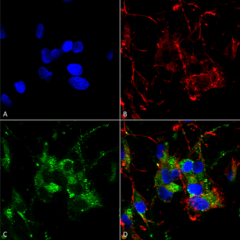
Immunocytochemistry/Immunofluorescence analysis using Mouse Anti-HCN2 Monoclonal Antibody, Clone S71. Tissue: Neuroblastoma cells (SH-SY5Y). Species: Human. Fixation: 4% PFA for 15 min. Primary Antibody: Mouse Anti-HCN2 Monoclonal Antibody at 1:50 for overnight at 4°C with slow rocking. Secondary Antibody: AlexaFluor 488 at 1:1000 for 1 hour at RT. Counterstain: Phalloidin-iFluor 647 (red) F-Actin stain; Hoechst (blue) nuclear stain at 1:800, 1.6mM for 20 min at RT. (A) Hoechst (blue) nuclear stain. (B) Phalloidin-iFluor 647 (red) F-Actin stain. (C) HCN2 Antibody (D) Composite.

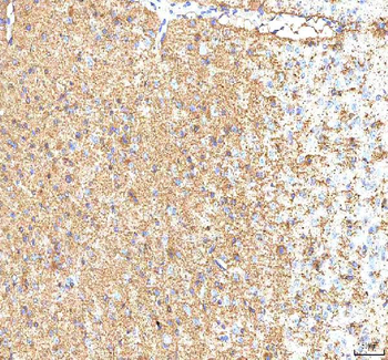
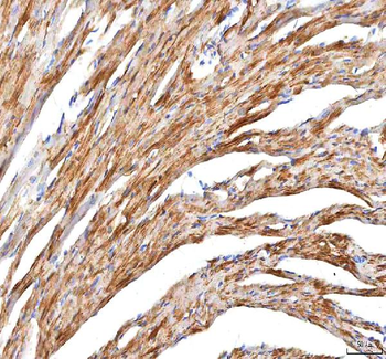
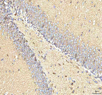
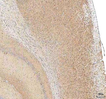
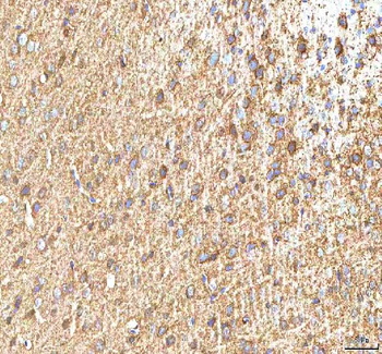
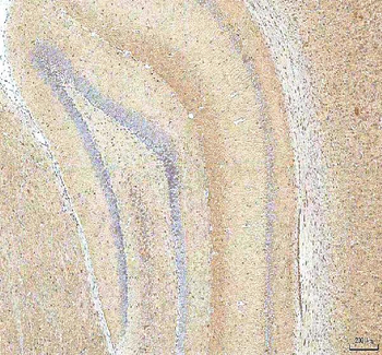
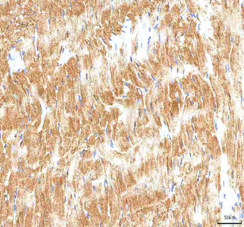
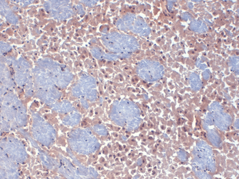
Immunohistochemistry analysis using Mouse Anti-HCN2 Monoclonal Antibody, Clone S71. Tissue: frozen brain section. Species: mouse. Fixation: 10% Formalin Solution for 12-24 hours at RT. Primary Antibody: Mouse Anti-HCN2 Monoclonal Antibody at 1:1000 for 1 hour at RT. Secondary Antibody: HRP/DAB Detection System: Biotinylated Goat Anti-Mouse, Streptavidin Peroxidase, DAB Chromogen (brown) for 30 minutes at RT. Counterstain: Mayer Hematoxylin (purple/blue) nuclear stain at 250-500 μl for 5 minutes at RT.
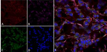
Immunocytochemistry/Immunofluorescence analysis using Mouse Anti-HCN2 Monoclonal Antibody, Clone S71-37. Tissue: Differentiated SH-SY5Y. Species: Human. Primary Antibody: Mouse Anti-HCN2 Monoclonal Antibody at 1:100. Secondary Antibody: AlexaFluor 488. Counterstain: phalloidin (Alexa 647, red), beta tubulin (Anti-beta III Tubulin Ab, Alexa 555, magenta) Hoechst (blue). (A) Phalloidin (B) Anti-beta III Tubulin Ab. (C) HCN2 Antibody. (D) Hoechst (E) Composite.

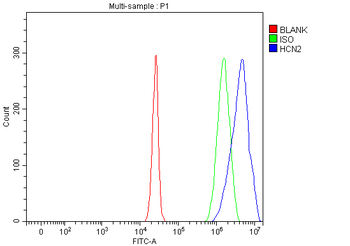
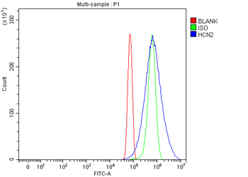
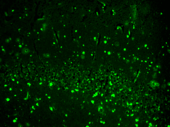
Immunohistochemistry analysis using Mouse Anti-HCN2 Monoclonal Antibody, Clone S71. Tissue: hippocampus. Species: Human. Fixation: Bouin's Fixative and paraffin-embedded. Primary Antibody: Mouse Anti-HCN2 Monoclonal Antibody at 1:100 for 1 hour at RT. Secondary Antibody: FITC Goat Anti-Mouse (green) at 1:50 for 1 hour at RT.

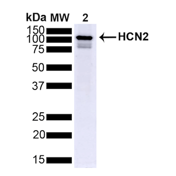
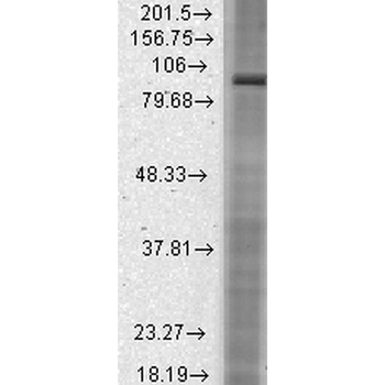
Western Blot analysis of Mouse Brain showing detection of ~95 kDa HCN2 protein using Mouse Anti-HCN2 Monoclonal Antibody, Clone S71. Lane 1: MW Ladder. Lane 2: Mouse Brain (15 ug). Load: 15 ug. Block: 5% Skim Milk powder in TBST. Primary Antibody: Mouse Anti-HCN2 Monoclonal Antibody at 1:1000 for 2 hours at RT with shaking. Secondary Antibody: Goat anti-mouse IgG:HRP at 1:4000 for 1 hour at RT with shaking. Color Development: Chemiluminescent for HRP (Moss) for 5 min in RT. Predicted/Observed Size: ~95 kDa.

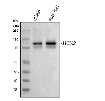
Western Blot analysis of Rat brain membrane lysate showing detection of HCN2 protein using Mouse Anti-HCN2 Monoclonal Antibody, Clone S71. Load: 15 μg. Block: 1.5% BSA for 30 minutes at RT. Primary Antibody: Mouse Anti-HCN2 Monoclonal Antibody at 1:1000 for 2 hours at RT. Secondary Antibody: Sheep Anti-Mouse IgG: HRP for 1 hour at RT.
Quick Database Links
UniProt Details
− No UniProt data available
NCBI Gene Details
− No NCBI Gene data available
NCBI Reference Sequences
−Associated Accession Numbers
Curated reference sequences for the gene transcript and protein product| Protein | NP_446136.1 |
|---|
Documents Download
Datasheet
Product Information
Request a Document
Protocol Information
WB
Western Blot (IB, immunoblot)
IHC
Immunohistochemistry
IF
Immunofluorescence
ICC
Immunocytochemistry
IP
Immunoprecipitation
HCN2 Antibody (orb67395)

- 0.0
Based on 0 reviews
Participating in our Biorbyt product reviews program enables you to support fellow scientists by sharing your firsthand experience with our products.
Login to Submit a ReviewAvailable Sizes
Select a size below
Choose Conjugation or Carrier Free Version
Free Secondary Antibody (20 ul)0/0
Please add an antibody product to your cart first.