You have no items in your shopping cart.
Description
Research Area
Metabolism Research
Images & Validation
−Item 1 of 4
| Tested Applications | ELISA, IF, IHC, WB |
|---|---|
| Dilution Range | Peptide ELISA: antibody detection limit dilution 1:64000. Western blot: Approx. 75kDa band observed in lysates of cell lines HEK293 and NIH3T3 and in Rat Heart lysates, and approx. 80kDa in Human and Mouse Heart and in Human Skeletal Muscle lysates (calculated MW of 79.5kDa according to Human NP_004997.4 and Rat NP_001005550.1, 75.4kDa according to Human NP_001186910.1, and 79.8kDa according to Mouse NP_001153510.1). Recommended concentration: 0.1-0.5µg/ml. Primary incubation 1 hour at room temperature. IHC: Paraffin embedded Human Heart. Recommended concentration: 5µg/ml. Immunofluorescence: Strong expression of the protein seen in U2OS cells. Recommended concentration: 10µg/ml. Flow Cytometry: Flow cytometric analysis of HEK293 cells. Recommended concentration: 10ug/ml. |
| Reactivity | Human, Mouse |
| Predicted Reactivity | Bovine, Canine, Rat |
Key Properties
−| Host | Goat |
|---|---|
| Clonality | Polyclonal |
| Target | NDUFS1 |
| Protein Sequence | TEKSATYVNTEGR |
| Molecular Weight | 79.5 |
| Purification | Purified from goat serum by ammonium sulphate precipitation followed by antigen affinity chromatography using the immunizing peptide. |
| Conjugation | Unconjugated |
Storage & Handling
−| Storage | Maintain refrigerated at 2-8°C for up to 2 weeks. For long term storage store at -20°C in small aliquots to prevent freeze-thaw cycles. |
|---|---|
| Buffer/Preservatives | Supplied at 0.5 mg/ml in Tris saline, 0.02% sodium azide, pH 7.3 with 0.5% bovine serum albumin. Aliquot and store at -20°C. Minimize freezing and thawing. |
| Expiration Date | 12 months from date of receipt. |
| Disclaimer | For research use only |
Alternative Names
−anti CI-75Kd antibody, anti complex I, mitochondrial respirato antibody, anti MGC26839 antibody, anti NADH dehydrogenase (ubiquinone) Fe-S protein 1, 75kDa antibody, anti NADH dehydrogenase (ubiquinone) Fe-S protein 1, 75kDa (NADH-coenzyme Q reductase) antibody, anti NADH-coenzyme Q reductase antibody, anti OTTHUMP00000206345 antibody, anti PRO1304 antibody, anti NDUFS1 antibody
Similar Products
−- Item 1 of 1
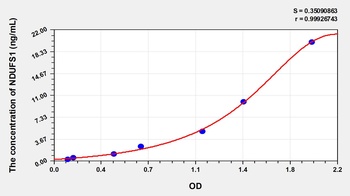
Human NADH Dehydrogenase Ubiquinone Fe-S Protein 1 (NDUFS1) ELISA Kit [orb778524]
Human
0.32-20 ng/mL
0.132 ng/mL
48 T, 96 T - Item 1 of 5
NDUFS1 Rabbit Polyclonal Antibody [orb556883]
ICC, IHC-P, WB
Human, Mouse, Rat
Rabbit
Polyclonal
Unconjugated
100 μl - Item 1 of 3
NDUFS1 Antibody [orb521260]
ELISA, IHC, WB
Human, Mouse, Rat
Rabbit
Polyclonal
Unconjugated
50 μl, 100 μl - Item 1 of 3
NDUFS1 Antibody [orb521261]
ELISA, IHC, WB
Human, Mouse, Rat
Rabbit
Polyclonal
Unconjugated
50 μl, 100 μl - Item 1 of 3


Quality Guarantee
Explore bioreagents carefree to elevate your research. All our products are rigorously tested for performance. If a product does not perform as described on its datasheet, our scientific support team will provide expert troubleshooting, a prompt replacement, or a refund. For full details, please see our Terms & Conditions and Buying Guide. Contact us at [email protected].
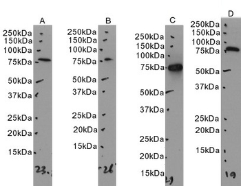
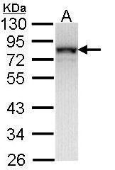
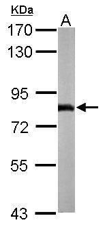
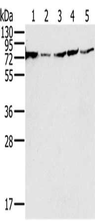
Primary incubation 1 hour at room temperature. Images A, B, C: Human Heart, Human Skeletal Muscle, Rat Heart lysate at primary Ab concentration 0.3 ug/ml. Image D: Mouse Heart lysate at primary Ab concentration 0.1 ug/ml (Loaded 35 µg protein in RIPA buffer, per lane). Detected by chemiluminescence.
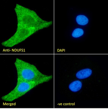
Immunofluorescence analysis of paraformaldehyde fixed U2OS cells, permeabilized with 0.15% Triton. Primary incubation 1hr (10 ug/ml) followed by Alexa Fluor 488 secondary antibody (2 ug/ml), showing mkitochondria/cytoplasmic staining. The nuclear stain is DAPI (blue). Negative control: Unimmunized goat IgG (10 ug/ml) followed by Alexa Fluor 488 secondary antibody (2 ug/ml).
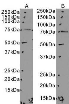
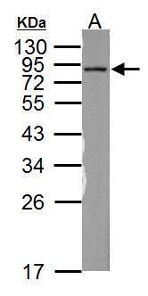
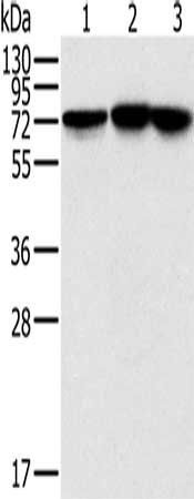
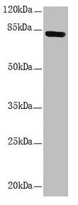
Primary incubation 1 hour at room temperature. Images A, B: HEK293, NIH3T3 cell lysate at primary Ab concentration 0.5 ug/ml. (Loaded 35 µg protein in RIPA buffer, per lane). Detected by chemiluminescence.
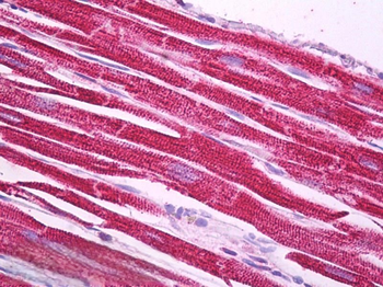
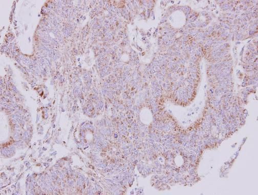
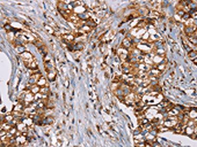
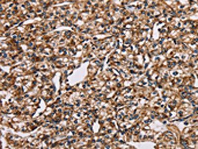
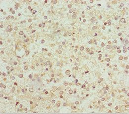
5 µg/ml staining of paraffin embedded Human Heart. Steamed antigen retrieval with citrate buffer pH6, AP-staining.
Documents Download
Datasheet
Product Information
Request a Document
Protocol Information
WB
Western Blot (IB, immunoblot)
IHC
Immunohistochemistry
IF
Immunofluorescence
ELISA
Enzyme-linked Immunosorbent Assay (EIA)
NDUFS1 Antibody (orb20594)

- 0.0
Based on 0 reviews
Participating in our Biorbyt product reviews program enables you to support fellow scientists by sharing your firsthand experience with our products.
Login to Submit a ReviewAvailable Sizes
Select a size below
Choose Conjugation or Carrier Free Version
Free Secondary Antibody (20 ul)0/0
Please add an antibody product to your cart first.