You have no items in your shopping cart.
Description
Research Area
Epigenetics & Chromatin
Images & Validation
−Item 1 of 4
| Tested Applications | ChIP, ELISA, IF, WB |
|---|---|
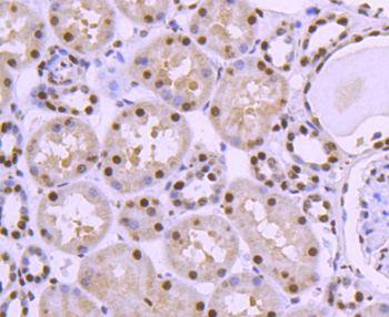
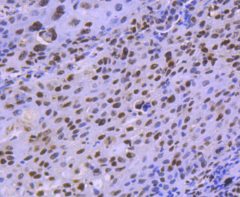
| Dilution Range | ELISA: 1:4000, WB: 0.1-2 μg/ml, IHC-P: 10 μg/ml |
| Reactivity | Human, Mouse |
| Predicted Reactivity | Canine, Rat |
| Application Notes |
Key Properties
−| Clonality | Polyclonal |
|---|---|
| Target | Histone Deacetylase 1 |
| Protein Sequence | KPEAKGVKEEVK |
| Molecular Weight | 55.1 |
| Purification | Purified from goat serum by ammonium sulphate precipitation followed by antigen affinity chromatography using the immunizing peptide. |
| Conjugation | Unconjugated |
Storage & Handling
−| Storage | Maintain refrigerated at 2-8°C for up to 2 weeks. For long term storage store at -20°C in small aliquots to prevent freeze-thaw cycles. |
|---|---|
| Buffer/Preservatives | Supplied at 0.5 mg/ml in Tris saline, 0.02% sodium azide, pH 7.3 with 0.5% bovine serum albumin. Aliquot and store at -20°C. Minimize freezing and thawing. |
| Expiration Date | 12 months from date of receipt. |
| Disclaimer | For research use only |
Alternative Names
−anti HDAC1 antibody, anti histone deacetylase 1 antibody, anti DKFZp686H12203 antibody, anti GON-10 antibody, anti HD1 antibody, anti RPD3 antibody, anti RPD3L1 antibody, anti reduced potassium dependency, yeast homolog-like 1 antibody
Similar Products
−- Item 1 of 8
HDAC8 Recombinant Rabbit Monoclonal Antibody [orb559101]
FC, ICC, WB
Mouse, Rat
Human
Rabbit
Recombinant
Unconjugated
50 μl, 100 μl - Item 1 of 4
HDAC1 Rabbit Polyclonal Antibody [orb10812]
FC, IF, IHC-Fr, IHC-P, WB
Bovine, Canine, Guinea pig, Porcine, Rat, Sheep
Human, Mouse
Rabbit
Polyclonal
Unconjugated
50 μl, 100 μl, 200 μl - Item 1 of 5
HDAC3 Antibody [orb242882]
ChIP, ELISA, IF, IHC, WB
Human, Mouse
Rabbit
Polyclonal
Unconjugated
100 μg, 50 μg - Item 1 of 4
HDAC1 rabbit pAb Antibody [orb765373]
ELISA, IF, IHC, WB
Human, Mouse, Rat
Polyclonal
Unconjugated
50 μl, 100 μl - Item 1 of 1


Quality Guarantee
Explore bioreagents carefree to elevate your research. All our products are rigorously tested for performance. If a product does not perform as described on its datasheet, our scientific support team will provide expert troubleshooting, a prompt replacement, or a refund. For full details, please see our Terms & Conditions and Buying Guide. Contact us at [email protected].

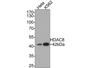
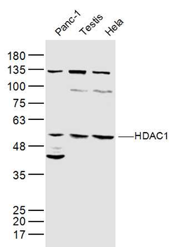
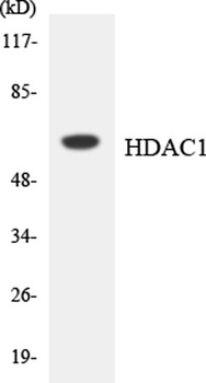
Western blot analysis of HeLa lysate (35µg protein in RIPA buffer) using HDAC1 antibody.
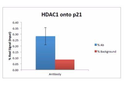
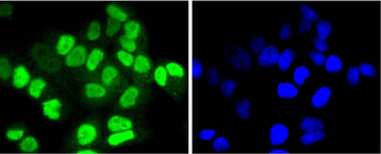
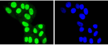
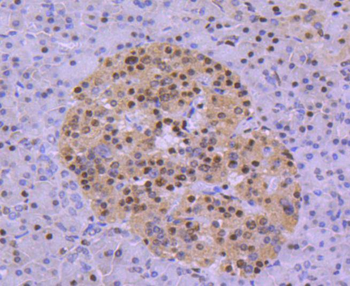
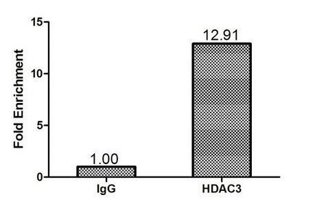
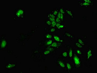
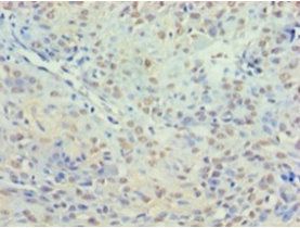
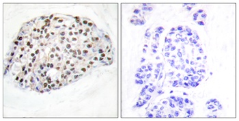
Chromatin immunoprecipitation analysis of using HDAC1 antibody
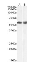
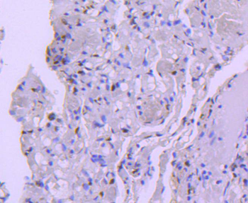
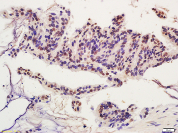
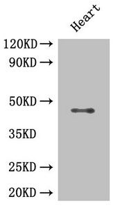
Western blot analysis of Human Duodenum (A) and Lymph Node (B) lysate using HDAC1 antibody.
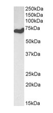
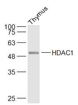
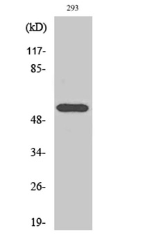
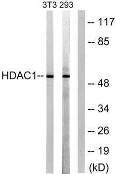
Western blot analysis of NIH3T3 lysate using HDAC1 antibody.
Documents Download
Datasheet
Product Information
Request a Document
Protocol Information
WB
Western Blot (IB, immunoblot)
IF
Immunofluorescence
ELISA
Enzyme-linked Immunosorbent Assay (EIA)
ChIP
Chromatin Immunoprecipitation
Histone Deacetylase 1 Antibody (orb12395)

- 0.0
Based on 0 reviews
Participating in our Biorbyt product reviews program enables you to support fellow scientists by sharing your firsthand experience with our products.
Login to Submit a ReviewAvailable Sizes
Select a size below
Choose Conjugation or Carrier Free Version
Free Secondary Antibody (20 ul)0/0
Please add an antibody product to your cart first.