You have no items in your shopping cart.
Description
Research Area
Immunology & Inflammation; Cell Biology
Images & Validation
−Item 1 of 6
| Tested Applications | ELISA, IF, WB |
|---|---|
| Dilution Range | ELISA: 1:128000, WB: 0.3-1 μg/ml |
| Reactivity | Human |
| Predicted Reactivity | Canine |
| Application Notes |
Key Properties
−| Clonality | Polyclonal |
|---|---|
| Target | CD47 |
| Protein Sequence | SNQKTIQPPRK |
| Molecular Weight | 35.2; 33.2 |
| Purification | Purified from goat serum by ammonium sulphate precipitation followed by antigen affinity chromatography using the immunizing peptide. |
| Conjugation | Unconjugated |
Storage & Handling
−| Storage | Maintain refrigerated at 2-8°C for up to 2 weeks. For long term storage store at -20°C in small aliquots to prevent freeze-thaw cycles. |
|---|---|
| Buffer/Preservatives | Supplied at 0.5 mg/ml in Tris saline, 0.02% sodium azide, pH 7.3 with 0.5% bovine serum albumin. Aliquot and store at -20°C. Minimize freezing and thawing. |
| Expiration Date | 12 months from date of receipt. |
| Disclaimer | For research use only |
Alternative Names
−CD47 antibody, CD47 molecule antibody, IAP antibody, MER6 antibody, OA3 antibody, CD47 antigen (Rh-related antigen, integrin-associated signal transducer) antibody, CD47 glycoprotein antibody, Rh-related antigen antibody, antigen identified by monoclonal antibody 1D8 antibody, antigenic surface determinant protein OA3 antibody, integrin associ antibody
Similar Products
−- Item 1 of 10
CD47 Rabbit Polyclonal Antibody [orb389308]
ICC, IF, IHC-P, WB
Guinea pig, Mouse, Rat
Rabbit
Polyclonal
Unconjugated
100 μg - Item 1 of 5
CD47 Rabbit Polyclonal Antibody [orb4838]
FC, WB
Rabbit
Human, Mouse, Rat
Rabbit
Polyclonal
Unconjugated
50 μl, 100 μl, 200 μl - Item 1 of 6
- Item 1 of 7
- Item 1 of 6


Quality Guarantee
Explore bioreagents carefree to elevate your research. All our products are rigorously tested for performance. If a product does not perform as described on its datasheet, our scientific support team will provide expert troubleshooting, a prompt replacement, or a refund. For full details, please see our Terms & Conditions and Buying Guide. Contact us at [email protected].
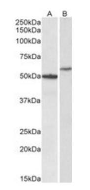
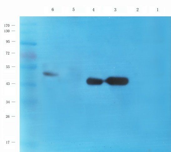
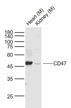
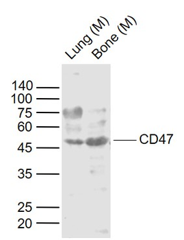
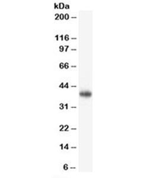
Western blot analysis of Human Hippocampus (Lane 1) and HeLa (Lane 2) lysates using CD47 antibody

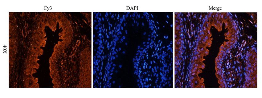
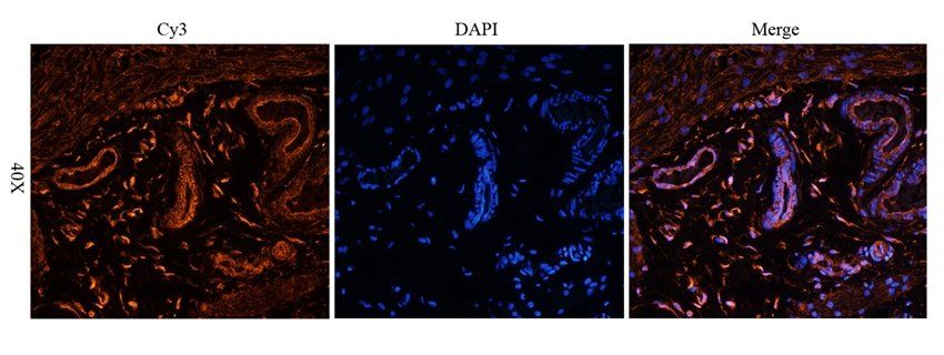
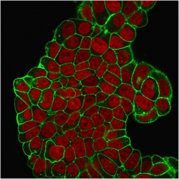
Immunofluorescence analysis of paraformaldehyde fixed A431 cells, permeabilized with 0.15% Triton. Primary incubation 1 hr (10 µg/mL) followed by Alexa Fluor 488 secondary antibody (2 µg/mL), showing cytoplasmic and membrane staining. The nuclear stain is DAPI (blue). Negative control: Unimmunized goat IgG (10 µg/mL) followed by Alexa Fluor 488 secondary antibody (2 µg/mL).
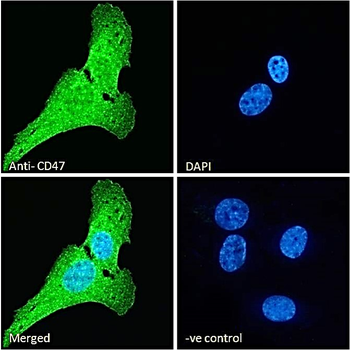
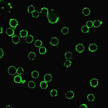
Immunofluorescence analysis of paraformaldehyde fixed U2OS cells, permeabilized with 0.15% Triton. Primary incubation 1 hr (10 µg/mL) followed by Alexa Fluor 488 secondary antibody (2 µg/mL), showing membrane, cytoplasmic and some nuclear staining. The nuclear stain is DAPI (blue). Negative control: Unimmunized goat IgG (10 µg/mL) followed by Alexa Fluor 488 secondary antibody (2 µg/mL).
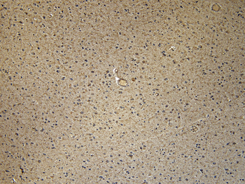
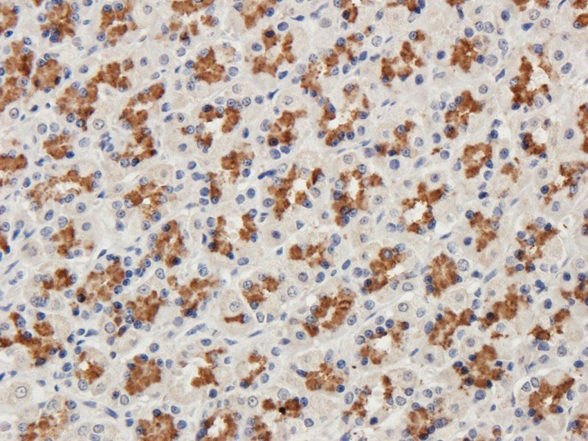
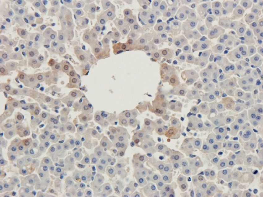
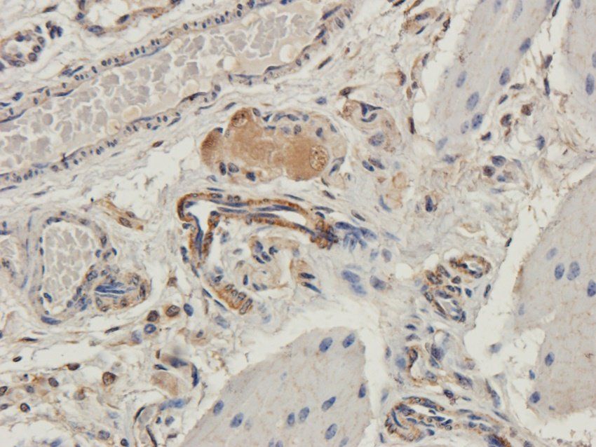
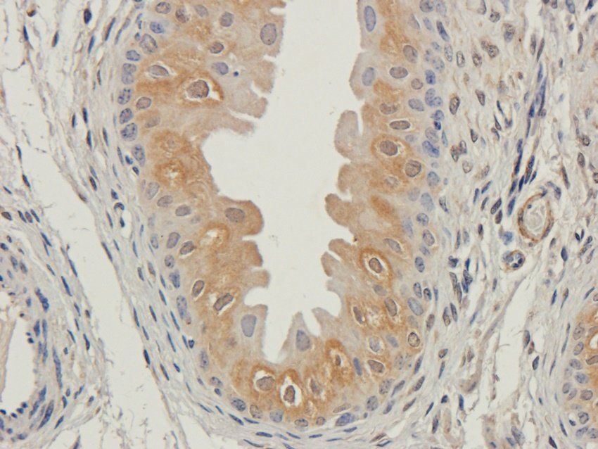
7 µg/mL staining of paraffin embedded Human Cortex. Heat induced antigen retrieval with citrate buffer pH 6, HRP-staining.
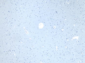
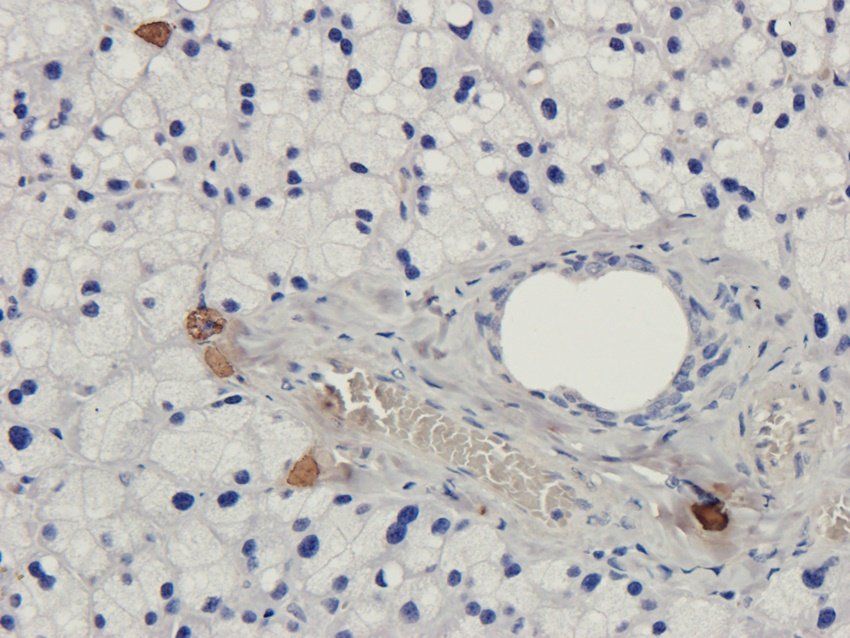
Negative Control showing staining of paraffin embedded Human Cortex, with no primary antibody.
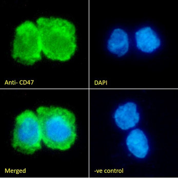
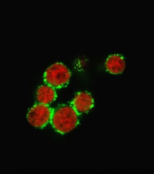
Immunofluorescence analysis of paraformaldehyde fixed THP-1 cells immobilized on ShifixTM coverslip, permeabilized with 0.15% Triton. Primary incubation 1 hr (10 µg/mL) followed by Alexa Fluor 488 secondary antibody (2 µg/mL), showing membrane and cytoplasmic staining. The nuclear stain is DAPI (blue). Negative control: Unimmunized goat IgG (10 µg/mL) followed by Alexa Fluor 488 secondary antibody (2 µg/mL).
Documents Download
Datasheet
Product Information
Request a Document
Protocol Information
WB
Western Blot (IB, immunoblot)
IF
Immunofluorescence
ELISA
Enzyme-linked Immunosorbent Assay (EIA)
CD47 Antibody (orb181434)

- 0.0
Based on 0 reviews
Participating in our Biorbyt product reviews program enables you to support fellow scientists by sharing your firsthand experience with our products.
Login to Submit a ReviewAvailable Sizes
Select a size below
Choose Conjugation or Carrier Free Version
Free Secondary Antibody (20 ul)0/0
Please add an antibody product to your cart first.