You have no items in your shopping cart.
Description
Research Area
Signal Transduction
Images & Validation
−Item 1 of 3
| Tested Applications | ELISA, FC, IF, WB |
|---|---|
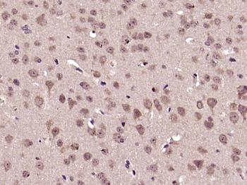
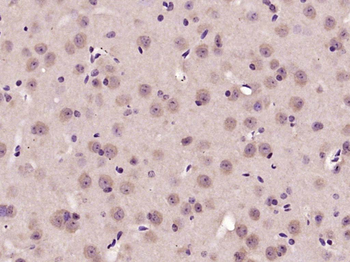
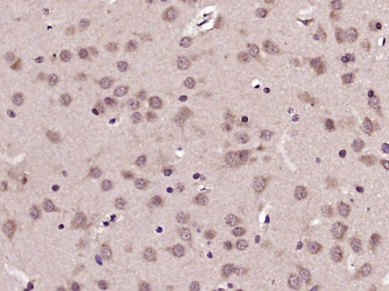
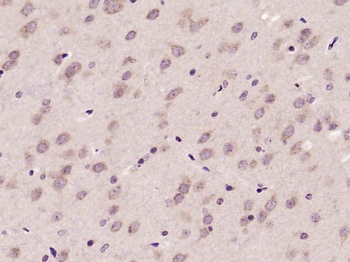
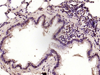
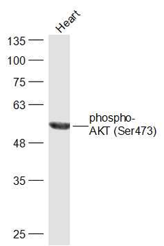
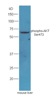
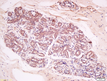
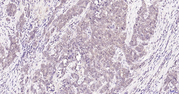
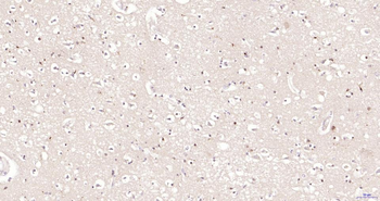
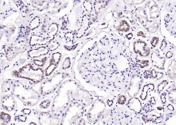
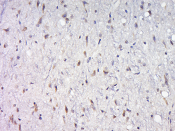
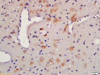
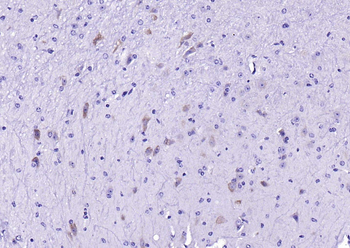
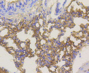
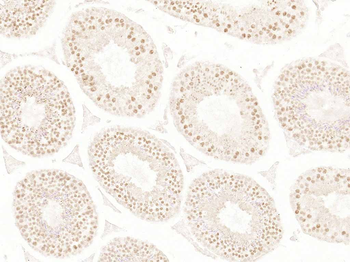
| Dilution Range | ELISA: 1:32000, WB: 0.1-0.3ug/ml, IHC-P: 10 μg/ml |
| Reactivity | Human |
| Predicted Reactivity | Mouse |
| Application Notes |
Key Properties
−| Clonality | Polyclonal |
|---|---|
| Target | AKT1 |
| Protein Sequence | QDVDQREAPLN |
| Molecular Weight | 55.7 |
| Purification | Purified from goat serum by ammonium sulphate precipitation followed by antigen affinity chromatography using the immunizing peptide. |
| Conjugation | Unconjugated |
Storage & Handling
−| Storage | Maintain refrigerated at 2-8°C for up to 2 weeks. For long term storage store at -20°C in small aliquots to prevent freeze-thaw cycles. |
|---|---|
| Buffer/Preservatives | Supplied at 0.5 mg/ml in Tris saline, 0.02% sodium azide, pH 7.3 with 0.5% bovine serum albumin. Aliquot and store at -20°C. Minimize freezing and thawing. |
| Expiration Date | 12 months from date of receipt. |
| Disclaimer | For research use only |
Alternative Names
−AKT1; v-akt murine thymoma viral oncogene homolog 1 ; AKT; MGC99656; PKB; PRKBA; RAC; RAC-ALPHA ; RAC-alpha serine/threonine-protein kinase; murine thymoma viral (v-akt) oncogene homolog-1; protein kinase B; rac protein kinase alpha
Similar Products
−- Item 1 of 11
Phospho-AKT (Ser473) Rabbit Polyclonal Antibody [orb11293]
WB
Bovine, Canine, Gallus, Mouse, Porcine, Rabbit, Rat, Sheep, Zebrafish
Human
Rabbit
Polyclonal
Unconjugated
50 μl, 100 μl, 200 μl - Item 1 of 9
AKT1 Antibody [orb344403]
ELISA, IF, IHC, Multiplex Assay, WB
Human, Monkey, Mouse, Rat
Mouse
Monoclonal
Unconjugated
100 μg - Item 1 of 11
Phospho-Akt (Thr450) Recombinant Rabbit Monoclonal Antibody [orb559195]
IF, IHC-Fr, IHC-P
Mouse, Rat
Human, Mouse, Rat
Rabbit
Recombinant
Unconjugated
50 μl, 100 μl, 25 μl - Item 1 of 8
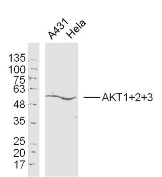
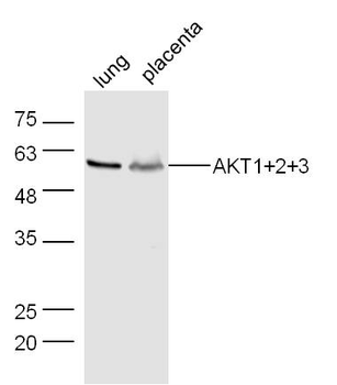
AKT1+2+3 Rabbit Polyclonal Antibody [orb155629]
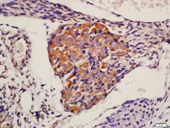
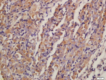
FC, IF, IHC-Fr, IHC-P, WB
Bovine, Canine, Gallus, Porcine, Rabbit, Sheep
Human, Mouse, Rat
Rabbit
Polyclonal
Unconjugated
50 μl, 100 μl, 200 μl - Item 1 of 8
Phospho-Akt1 (Ser473) Recombinant Rabbit Monoclonal Antibody [orb526646]
WB
Mouse, Rat, Zebrafish
Human
Rabbit
Recombinant
Unconjugated
50 μl, 100 μl, 25 μl


Quality Guarantee
Explore bioreagents carefree to elevate your research. All our products are rigorously tested for performance. If a product does not perform as described on its datasheet, our scientific support team will provide expert troubleshooting, a prompt replacement, or a refund. For full details, please see our Terms & Conditions and Buying Guide. Contact us at [email protected].
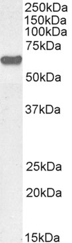
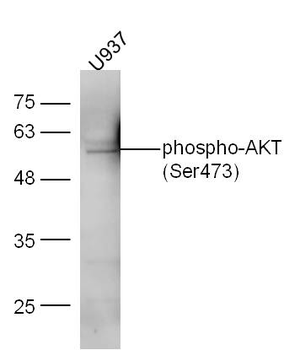
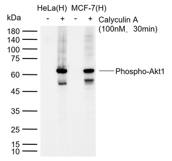
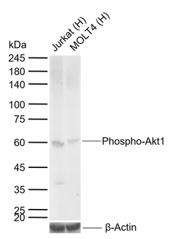
0.3 μg/ml staining of MCF7 cell lysate (35 μg protein in RIPA buffer). Detected by chemiluminescence.
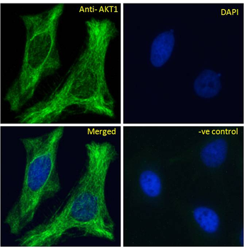
Immunofluorescence analysis of paraformaldehyde fixed HeLa cells, permeabilized with 0.15% Triton. Primary incubation 1hr (10 μg/ml) followed by Alexa Fluor 488 secondary antibody (2 μg/ml), showing Microtubule/cytoplasmic staining. The nuclear stain is DAPI (blue). Negative control: Unimmunized goat IgG (10 μg/ml) followed by Alexa Fluor 488 secondary antibody (2 μg/ml).
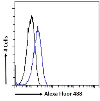
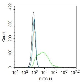
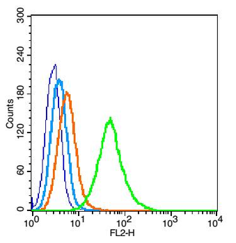
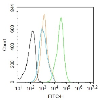
Flow cytometric analysis of paraformaldehyde fixed MCF7 cells (blue line), permeabilized with 0.5% Triton. Primary incubation 1hr (10 μg/ml) followed by Alexa Fluor 488 secondary antibody (1 μg/ml). IgG control: Unimmunized goat IgG (black line) followed by Alexa Fluor 488 secondary antibody.
Documents Download
Datasheet
Product Information
Request a Document
Protocol Information
WB
Western Blot (IB, immunoblot)
FC
Flow Cytometry
IF
Immunofluorescence
ELISA
Enzyme-linked Immunosorbent Assay (EIA)
AKT1 Antibody (orb422482)

- 0.0
Based on 0 reviews
Participating in our Biorbyt product reviews program enables you to support fellow scientists by sharing your firsthand experience with our products.
Login to Submit a ReviewAvailable Sizes
Select a size below
Choose Conjugation or Carrier Free Version
Free Secondary Antibody (20 ul)0/0
Please add an antibody product to your cart first.