You have no items in your shopping cart.
Featured
Description
Research Area
Cancer Research, Cardiovascular Research, Diabetes Research, Metabolism
Images & Validation
−Item 1 of 10
| Tested Applications | ICC, IF, IHC-Fr, IHC-P |
|---|---|
| Dilution Range | IHC-P=1:200-1000, IHC-F=1:200-1000, ICC/IF=1:50-200, IF=1:200-1000 |
| Reactivity | Human, Mouse, Rat |
| Predicted Reactivity | Mouse, Rat |
Related Conjugates & Formulations
−Key Properties
−| Antibody Type | Primary Antibody |
|---|---|
| Host | Rabbit |
| Clonality | Recombinant |
| Isotype | IgG |
| Clone No. | B4F7 |
| Immunogen | A synthesized peptide derived from human GLUT1 (450-492aa) |
| Target | SLC2A1 |
| Molecular Weight | 54 kDa |
| Purification | Affinity purified by Protein A |
| Conjugation | Unconjugated |
Storage & Handling
−| Storage | Maintain refrigerated at 2-8°C for up to 2 weeks. For long term storage store at -20°C in small aliquots to prevent freeze-thaw cycles. |
|---|---|
| Form/Appearance | Liquid |
| Buffer/Preservatives | 0.01M TBS (pH7.4) with 1% rAlbumin, 0.02% Proclin300 and 50% Glycerol. |
| Concentration | 1mg/ml |
| Expiration Date | 12 months from date of receipt. |
| Disclaimer | For research use only |
Alternative Names
−CSE; DYT17; DYT18; DYT9; EIG12; GLUT; GLUT-1; GLUT1; GLUT1DS; HTLVR; PED; SDCHCN; GT1; M100200; Rgsc200; GLUTB; GTG1; Gtg3; RATGTG1; GTR1_HUMAN; SLC2A1; Glucose transporter type 1, erythrocyte/brain (GLUT-1); HepG2 glucose transporter; GTR1_MOUSE; Glucose transporter type 1, erythrocyte/brain (GLUT-1 | GT1); GTR1_RAT; solute carrier family 2 member 1; human T-cell leukemia virus (I and II) receptor; choreoathetosis/spasticity, episodic (paroxysmal choreoathetosis/spasticity); solute carrier family 2 (facilitated glucose transporter), member 1; dystonia gene 18; dystonia gene 9
Similar Products
−
GLUT1 Rabbit Monoclonal Antibody [orb2989742]
IHC, WB
Human, Mouse, Rat
Rabbit
Monoclonal
Unconjugated
200 μl, 100 μl, 50 μl, 30 μlRabbit GLUT1, recombinant (monoclonal) Antibody [orb1882312]
IHC-P
Human
Rabbit
Monoclonal
Unconjugated
0.5 ml
Quality Guarantee
Explore bioreagents carefree to elevate your research. All our products are rigorously tested for performance. If a product does not perform as described on its datasheet, our scientific support team will provide expert troubleshooting, a prompt replacement, or a refund. For full details, please see our Terms & Conditions and Buying Guide. Contact us at [email protected].
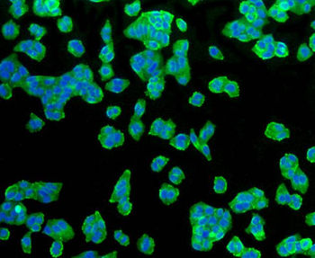
ICC staining of Glucose Transporter GLUT1 in Hela cells (green). Formalin fixed cells were permeabilized with 0.1% Triton X-100 in TBS for 10 minutes at room temperature and blocked with 1% Blocker BSA for 15 minutes at room temperature. Cells were probed with the primary antibody (orb612216, 1/50) for 1 hour at room temperature, washed with PBS. Alexa Fluor®488 Goat anti-Rabbit IgG was used as the secondary antibody at 1/1000 dilution. The nuclear counter stain is DAPI (blue).
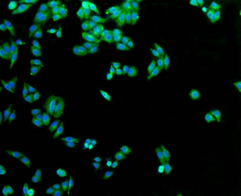
ICC staining of Glucose Transporter GLUT1 in HepG2 cells (green). Formalin fixed cells were permeabilized with 0.1% Triton X-100 in TBS for 10 minutes at room temperature and blocked with 1% Blocker BSA for 15 minutes at room temperature. Cells were probed with the primary antibody (orb612216, 1/50) for 1 hour at room temperature, washed with PBS. Alexa Fluor®488 Goat anti-Rabbit IgG was used as the secondary antibody at 1/1000 dilution. The nuclear counter stain is DAPI (blue).
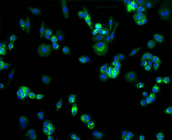
ICC staining of Glucose Transporter GLUT1 in MCF-7 cells (green). Formalin fixed cells were permeabilized with 0.1% Triton X-100 in TBS for 10 minutes at room temperature and blocked with 1% Blocker BSA for 15 minutes at room temperature. Cells were probed with the primary antibody (orb612216, 1/50) for 1 hour at room temperature, washed with PBS. Alexa Fluor®488 Goat anti-Rabbit IgG was used as the secondary antibody at 1/1000 dilution. The nuclear counter stain is DAPI (blue).
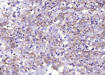
Paraformaldehyde-fixed, paraffin embedded (human endometrial carcinoma), Antigen retrieval by boiling in sodium citrate buffer (pH6.0) for 15 min, Block endogenous peroxidase by 3% hydrogen peroxide for 20 minutes, Blocking buffer (normal goat serum) at 37°C for 30 min, Antibody incubation with (GLUT1) Monoclonal Antibody, Unconjugated (orb612216) at 1:200 overnight at 4°C, followed by operating according to SP Kit (Rabbit) instructionsand DAB staining.
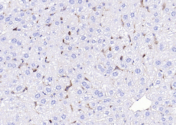
Paraformaldehyde-fixed, paraffin embedded (human liver), Antigen retrieval by boiling in sodium citrate buffer (pH6.0) for 15 min, Block endogenous peroxidase by 3% hydrogen peroxide for 20 minutes, Blocking buffer (normal goat serum) at 37°C for 30 min, Antibody incubation with (GLUT1) Monoclonal Antibody, Unconjugated (orb612216) at 1:200 overnight at 4°C, followed by operating according to SP Kit (Rabbit) instructionsand DAB staining.
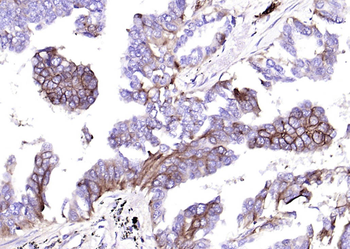
Paraformaldehyde-fixed, paraffin embedded (human lung carcinoma), Antigen retrieval by boiling in sodium citrate buffer (pH6.0) for 15 min, Block endogenous peroxidase by 3% hydrogen peroxide for 20 minutes, Blocking buffer (normal goat serum) at 37°C for 30 min, Antibody incubation with (GLUT1) Monoclonal Antibody, Unconjugated (orb612216) at 1:200 overnight at 4°C, followed by operating according to SP Kit (Rabbit) instructionsand DAB staining.
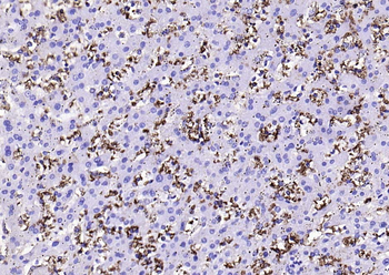
Paraformaldehyde-fixed, paraffin embedded (mouse liver), Antigen retrieval by boiling in sodium citrate buffer (pH6.0) for 15 min, Block endogenous peroxidase by 3% hydrogen peroxide for 20 minutes, Blocking buffer (normal goat serum) at 37°C for 30 min, Antibody incubation with (GLUT1) Monoclonal Antibody, Unconjugated (orb612216) at 1:200 overnight at 4°C, followed by operating according to SP Kit (Rabbit) instructionsand DAB staining.
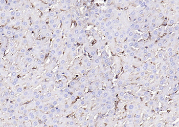
Paraformaldehyde-fixed, paraffin embedded (rat liver), Antigen retrieval by boiling in sodium citrate buffer (pH6.0) for 15 min, Block endogenous peroxidase by 3% hydrogen peroxide for 20 minutes, Blocking buffer (normal goat serum) at 37°C for 30 min, Antibody incubation with (GLUT1) Monoclonal Antibody, Unconjugated (orb612216) at 1:200 overnight at 4°C, followed by operating according to SP Kit (Rabbit) instructionsand DAB staining.
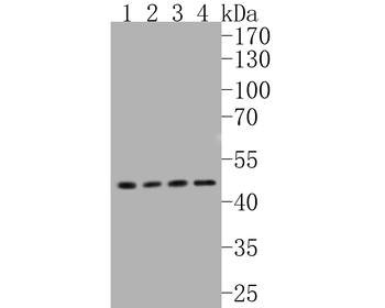
Western blot analysis of Glucose Transporter GLUT1 on different lysates. Proteins were transferred to a PVDF membrane and blocked with 5% BSA in PBS for 1 hour at room temperature. The primary antibody (orb612216, 1/500) was used in 5% BSA at room temperature for 2 hours. Goat Anti-Rabbit IgG - HRP Secondary Antibody at 1:5000 dilution was used for 1 hour at room temperature. Positive control: Lane 1: Hela cell lysate, Lane 2: SK-Br-3 cell lysate, Lane 3: NIH/3T3 cell lysate, Lane 4: HepG2 cell lysate.
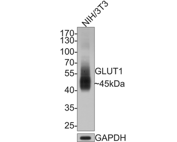
Western blot analysis of Glucose Transporter GLUT1 on NIH/3T3 cell lysates with Rabbit anti-Glucose Transporter GLUT1 antibody (orb612216) at 1/500 dilution. Lysates/proteins at 10 ug/lane. Predicted band size: 54 kDa, Observed band size: 45 kDa. Exposure time: 2 minutes. 10% SDS-PAGE gel.
Quick Database Links
Gene Symbol
SLC2A1
UniProt
UniProt Details
− No UniProt data available
Documents Download
Datasheet
Product Information
Request a Document
Protocol Information
IHC-P
Immunohistochemistry Paraffin
IHC-Fr
Immunohistochemistry Frozen
IF
Immunofluorescence
ICC
Immunocytochemistry
GLUT1 Recombinant Rabbit Monoclonal Antibody (orb612216)

- 0.0
Based on 0 reviews
Participating in our Biorbyt product reviews program enables you to support fellow scientists by sharing your firsthand experience with our products.
Login to Submit a ReviewAvailable Sizes
Select a size below
Free Secondary Antibody (20 ul)0/0
Please add an antibody product to your cart first.