You have no items in your shopping cart.
Featured
Description
Research Area
Neuroscience
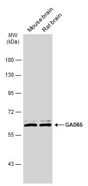
Images & Validation
−
Item 1 of 4
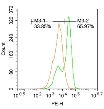
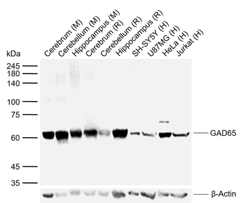
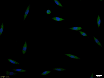
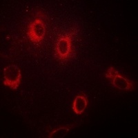
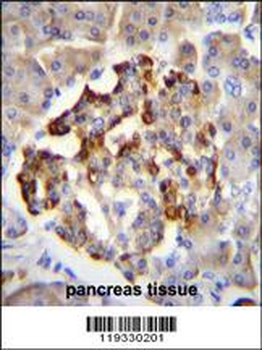
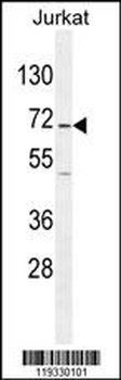
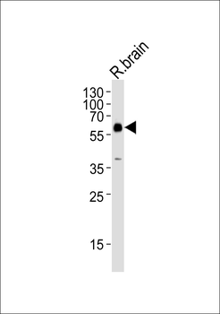
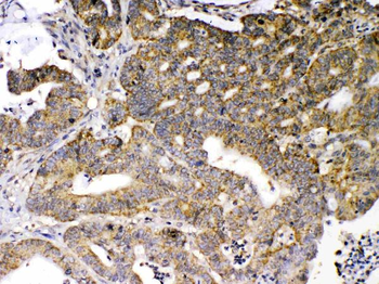
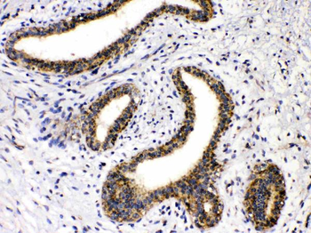
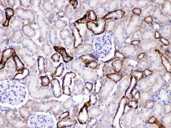
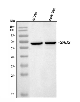
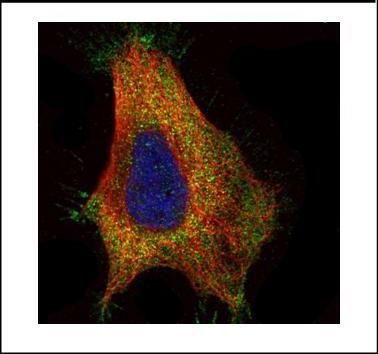
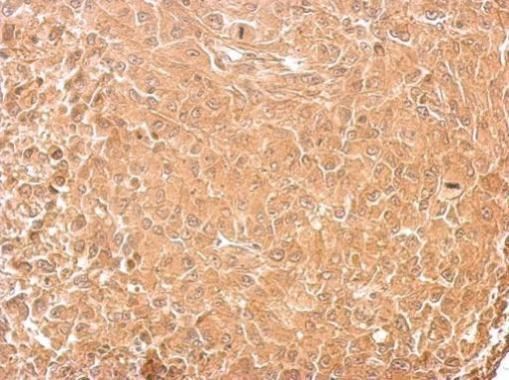
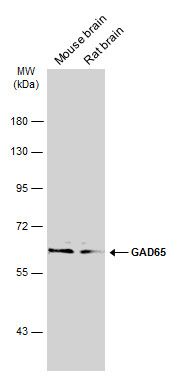
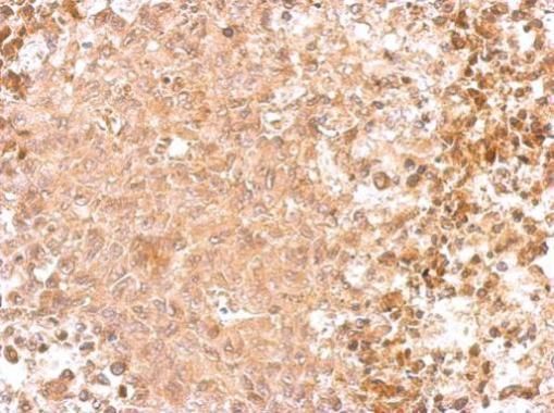
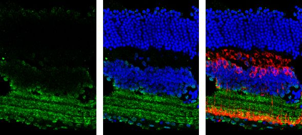
| Tested Applications | FC, ICC, WB |
|---|---|
| Dilution Range | WB=1:500-2000, ICC/IF=1:100-500, Flow-Cyt=1ug/Test |
| Reactivity | Human, Mouse, Rat |
| Predicted Reactivity | Bovine, Canine, Gallus, Porcine |
Related Conjugates & Formulations
−Key Properties
−| Antibody Type | Primary Antibody |
|---|---|
| Host | Rabbit |
| Clonality | Polyclonal |
| Isotype | IgG |
| Immunogen | KLH conjugated synthetic peptide derived from human GAD65 (501-585/585aa) |
| Target | GAD2 |
| Molecular Weight | 65 kDa |
| Purification | Affinity purified by Protein A |
| Conjugation | Unconjugated |
Storage & Handling
−| Storage | Maintain refrigerated at 2-8°C for up to 2 weeks. For long term storage store at -20°C in small aliquots to prevent freeze-thaw cycles. |
|---|---|
| Form/Appearance | Liquid |
| Buffer/Preservatives | 0.01M TBS (pH7.4) with 1% rAlbumin, 0.02% Proclin300 and 50% Glycerol. |
| Concentration | 1mg/ml |
| Expiration Date | 12 months from date of receipt. |
| Disclaimer | For research use only |
Alternative Names
−GAD65; 6330404F12Rik; GAD(65); Gad-2; DCE2_HUMAN; GAD2; 65 kDa glutamic acid decarboxylase (GAD-65); Glutamate decarboxylase 65 kDa isoform; 4.1.1.15; DCE2_MOUSE; DCE2_RAT;
Similar Products
−- Item 1 of 3
GAD1/2 Rabbit Polyclonal Antibody [orb213964]
IF, IHC, WB
Bovine, Canine, Human, Mouse, Porcine, Rat
Rabbit
Polyclonal
Unconjugated
30 μl, 100 μl, 200 μl, 50 μl - Item 1 of 4
GAD2 Antibody (Center) [orb1938434]
IF, IHC-P, WB
Mouse, Porcine, Rat
Human
Rabbit
Polyclonal
Unconjugated
100 μl - Item 1 of 4
GAD65/GAD2 Rabbit Polyclonal Antibody [orb389475]
IHC, WB
Human, Mouse, Rat
Rabbit
Polyclonal
Unconjugated
100 μg - Item 1 of 5
GAD65 Rabbit Polyclonal Antibody [orb556836]
ICC, IHC-P, WB
Human, Mouse, Rat
Rabbit
Polyclonal
Unconjugated
100 μl - Item 1 of 4
GAD65 Rabbit Polyclonal Antibody [orb555899]
ICC, IHC-Fr, IHC-P, WB
Human, Mouse, Rat
Rabbit
Polyclonal
Unconjugated
100 μl


Quality Guarantee
Explore bioreagents carefree to elevate your research. All our products are rigorously tested for performance. If a product does not perform as described on its datasheet, our scientific support team will provide expert troubleshooting, a prompt replacement, or a refund. For full details, please see our Terms & Conditions and Buying Guide. Contact us at [email protected].
Quick Database Links
Gene Symbol
GAD2
UniProt
UniProt Details
− No UniProt data available
Protocol Information
WB
Western Blot (IB, immunoblot)
FC
Flow Cytometry
ICC
Immunocytochemistry
Filter by Applications
Filter by Species
Fatemeh Bagheri Tadi et al. Astaxanthin improved carbamazepine efficacy and mood comorbidities in epileptic rats through GABAergic signaling pathway enhancement Research Square, (2026)
Applications
IF, WB
Reactivity
Rat
Janzadeh, Atousa et al. The effect of chondroitinase ABC and photobiomodulation therapy on neuropathic pain after spinal cord injury in adult male rats Physiol Behav, 227, 113141 (2020)