You have no items in your shopping cart.
Description
Research Area
Signal Transduction
Images & Validation
−Item 1 of 4
| Tested Applications | FC, IF, IHC-P, WB |
|---|---|
| Dilution Range | IF - 1:50-100, WB - 1:1000, IHC-P - 1:50-100, FC - 1:10-50 |
| Reactivity | Human, Mouse |
Key Properties
−| Antibody Type | Primary Antibody |
|---|---|
| Host | Rabbit |
| Clonality | Polyclonal |
| Isotype | Rabbit IgG |
| Immunogen | This FGFR1 antibody is generated from rabbits immunized with a KLH conjugated synthetic peptide between 19~48 amino acids from the N-terminal region of human FGFR1. Antigen Region: 19-48 aa. |
| Target | FGFR1 |
| Molecular Weight | 91868 Da |
| Conjugation | Unconjugated |
Storage & Handling
−| Storage | Maintain refrigerated at 2-8°C for up to 2 weeks. For long term storage store at -20°C in small aliquots to prevent freeze-thaw cycles |
|---|---|
| Form/Appearance | Purified polyclonal antibody supplied in PBS with 0.09% (W/V) sodium azide. This antibody is prepared by Saturated Ammonium Sulfate (SAS) precipitation followed by dialysis against PBS. |
| Expiration Date | 12 months from date of receipt. |
| Disclaimer | For research use only |
Alternative Names
−Fibroblast growth factor receptor 1, FGFR-1, Basic fibroblast growth factor receptor 1, BFGFR, bFGF-R-1, Fms-like tyrosine kinase 2, FLT-2, N-sam, Proto-oncogene c-Fgr, CD331, FGFR1, BFGFR, CEK, FGFBR, FLG, FLT2, HBGFR
Similar Products
−- Item 1 of 1
STAT3 Antibody (N-term) [orb106940]
WB
Mouse, Other, Porcine, Rat
Human
Rabbit
Polyclonal
Unconjugated
50 μl, 100 μl
Quality Guarantee
Explore bioreagents carefree to elevate your research. All our products are rigorously tested for performance. If a product does not perform as described on its datasheet, our scientific support team will provide expert troubleshooting, a prompt replacement, or a refund. For full details, please see our Terms & Conditions and Buying Guide. Contact us at [email protected].
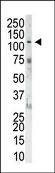
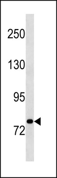
The anti-FGFR1 Pab is used in Western blot to detect FGFR1 in NIH-3T3 cell lysate.
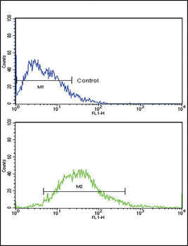
Flow cytometric analysis of MCF-7 cells using FGFR1 Antibody (N-term) (bottom histogram) compared to a negative control cell (top histogram). FITC-conjugated goat-anti-rabbit secondary antibodies were used for the analysis.
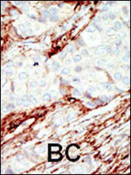
Formalin-fixed and paraffin-embedded human cancer tissue reacted with the primary antibody, which was peroxidase-conjugated to the secondary antibody, followed by DAB staining. This data demonstrates the use of this antibody for immunohistochemistry; clinical relevance has not been evaluated. BC = breast carcinoma; HC = hepatocarcinoma.
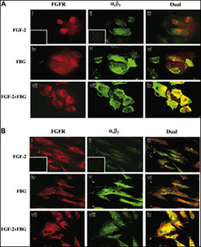
Colocalization of A1B3 and FGFR1 using IF. Confluent ECs (A) or HFFs (B) were treated with or without 100 ng/mL FGF-2 in the presence or absence of 10/mL fibrinogen. After 1 hour, cells were washed and fixed with 3.7% formaldehyde and stained using 10/mL FGFR1 and 7E3 antibody. FGFR is visualized as red fluorescence (i, iv, vii), A1B3 is visualized as green fluorescence (ii, v, viii), and colocalization of FGF-2 and fibrinogen receptors is shown as yellow fluorescence (iii, vi, ix). Insets represent the background staining for red (i) and green (ii) fluorescence. Bars represent 25.
Quick Database Links
Gene Symbol
FGFR1
UniProt
RefSeq (Protein):NP_001167535.1, NP_075593.1, NP_075598.2, NP_001167538.1, NP_056934.2, NP_001167536.1, NP_075594.1, NP_001167534.1, NP_001167537.1
UniProt Details
− No UniProt data available
NCBI Reference Sequences
−Associated Accession Numbers
Curated reference sequences for the gene transcript and protein productDocuments Download
Datasheet
Product Information
Request a Document
Protocol Information
WB
Western Blot (IB, immunoblot)
IHC-P
Immunohistochemistry Paraffin
FC
Flow Cytometry
IF
Immunofluorescence
FGFR1 Antibody (N-term) (orb1929052)

- 0.0
Based on 0 reviews
Participating in our Biorbyt product reviews program enables you to support fellow scientists by sharing your firsthand experience with our products.
Login to Submit a ReviewAvailable Sizes
Select a size below
Choose Conjugation or Carrier Free Version
Free Secondary Antibody (20 ul)0/0
Please add an antibody product to your cart first.