You have no items in your shopping cart.
Featured
Description
Images & Validation
−Item 1 of 8
| Tested Applications | ICC, IF, IHC-P, WB |
|---|---|
| Dilution Range | WB: 1:100-1500, IHC-P: 1:100-600, IF/ICC: 1:100-600 |
| Reactivity | Guinea pig, Mouse, Rat |
Key Properties
−| Host | Rabbit |
|---|---|
| Clonality | Polyclonal |
| Isotype | IgG |
| Immunogen | KLH conjugated synthetic peptide derived from human FFAR1. Please contact us for the exact immunogen sequence. The peptide is available as orb14118. |
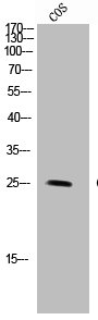
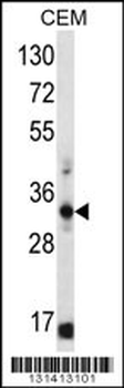
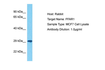
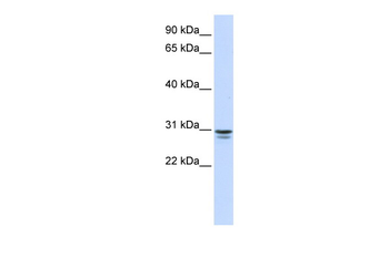
| Target | FFAR1 |
| Molecular Weight | 31 kDa |
| Purity | Polyclonal antibodies are purified by peptide affinity chromatography |
| Conjugation | Unconjugated |
Storage & Handling
−| Storage | Maintain refrigerated at 2-8°C for up to 2 weeks. For long term storage store at -20°C in small aliquots to prevent freeze-thaw cycles. |
|---|---|
| Form/Appearance | 10 mM PBS, 0.02% sodium azide |
| Concentration | - 100 μg (in 200 μl): 0.5 mg/ml- 200 μg (in 400 μl): 0.5 mg/ml |
| Expiration Date | 12 months from date of receipt. |
| Disclaimer | For research use only |
Similar Products
−- Item 1 of 4
GPR40 rabbit pAb Antibody [orb768492]
ELISA, IF, WB
Human, Monkey
Polyclonal
Unconjugated
50 μl, 100 μl - Item 1 of 3
- Item 1 of 1
- Item 1 of 2
FFAR1 Rabbit Polyclonal Antibody [orb331568]
WB
Bovine, Canine, Mouse, Porcine, Rat
Human
Rabbit
Polyclonal
Unconjugated
100 μl - Item 1 of 1
FFAR1 Rabbit Polyclonal Antibody [orb2951406]
ELISA, IHC, WB
Human
Rabbit
Polyclonal
Unconjugated
50 μg, 100 μg


Quality Guarantee
Explore bioreagents carefree to elevate your research. All our products are rigorously tested for performance. If a product does not perform as described on its datasheet, our scientific support team will provide expert troubleshooting, a prompt replacement, or a refund. For full details, please see our Terms & Conditions and Buying Guide. Contact us at [email protected].
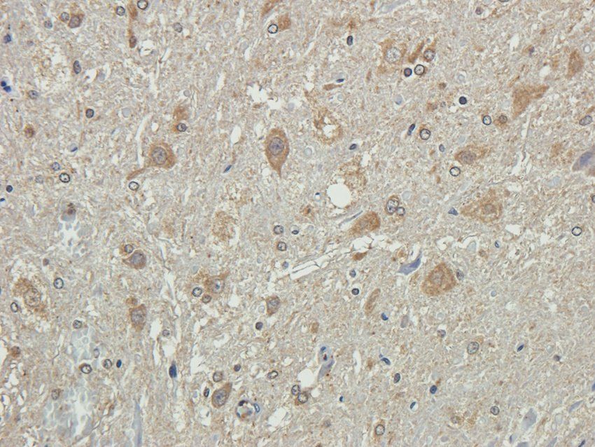
IHC-P image of rat spinal cord tissue using anti-FFAR1 (2.5 ug/ml)
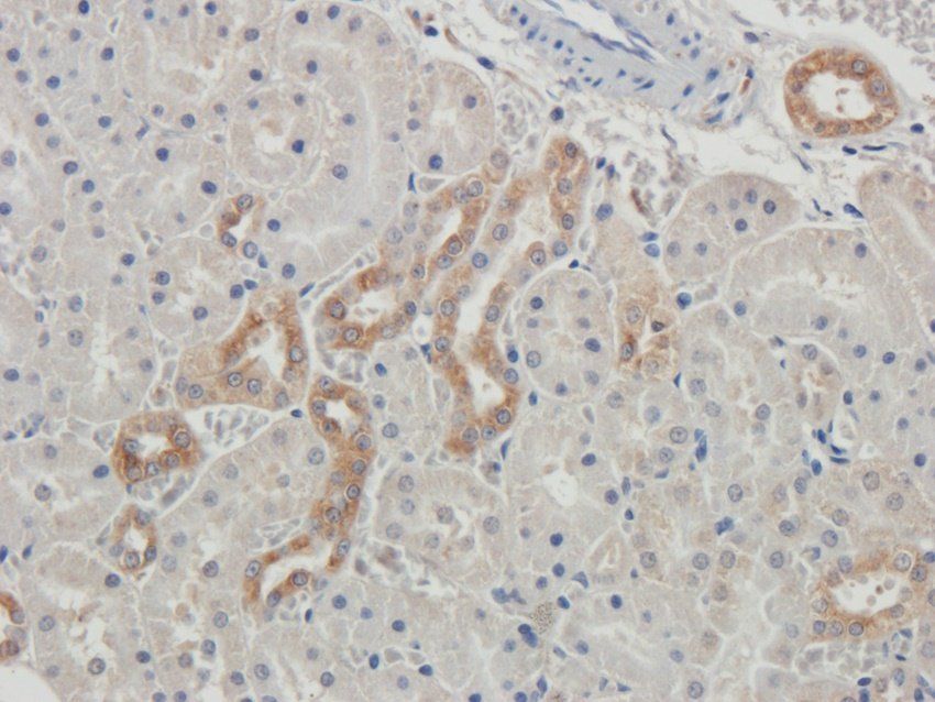
IHC-P staining of rat kidney tissue using anti-FFAR1 (2.5 ug/ml)
Immunohistochemical staining of paraffin embedded mouse ovary tissue using FFAR1 antibody (2.5 ug/ml)
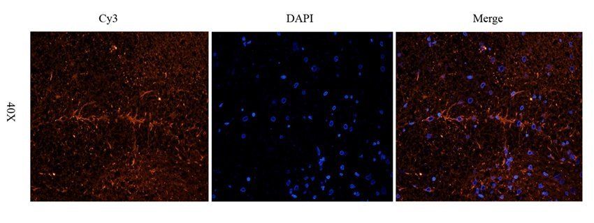
IF analysis of rat spinal cord tissue using anti-FFAR1 (2.5 ug/ml)
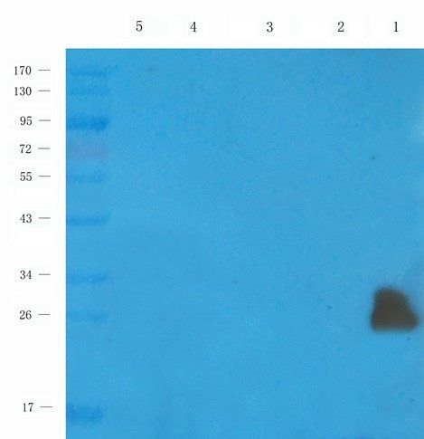
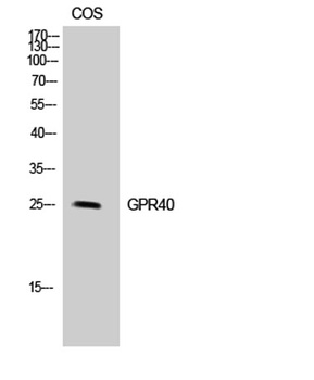
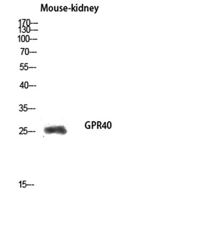
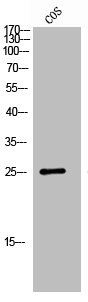
Western blot analysis of mouse pancreas (lane 1), rat ovary (lane 2), mouse spinal cord (lane 3), mouse brain (lane 4), mouse kidney (lane 5) using anti-FFAR1 (1 ug/ml)
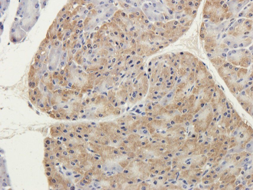
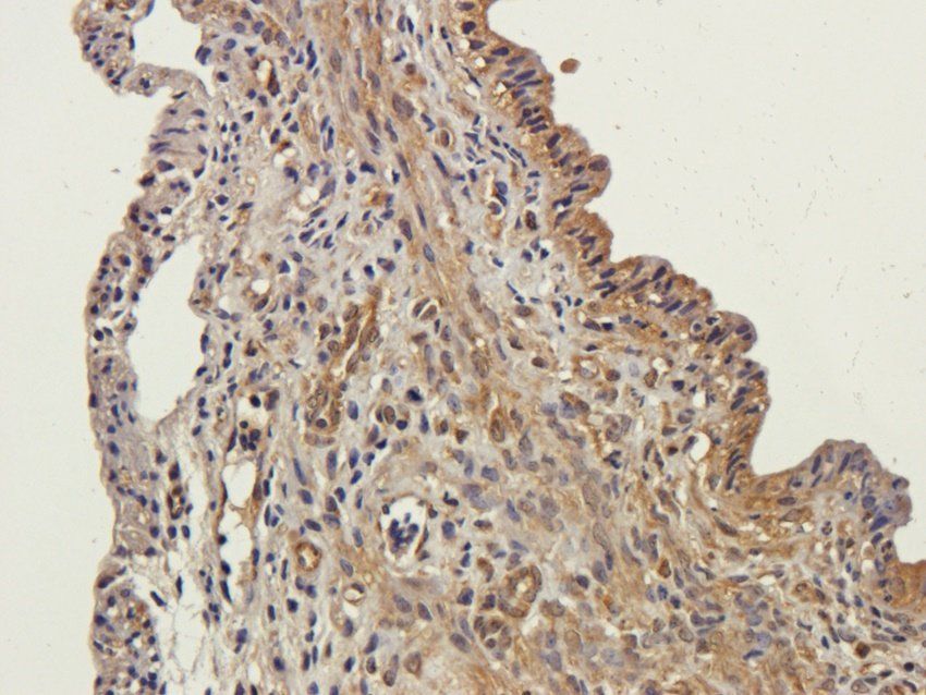
IHC-P staining of guinea pig pancreas tissue using anti-FFAR1 (2.5 ug/ml)
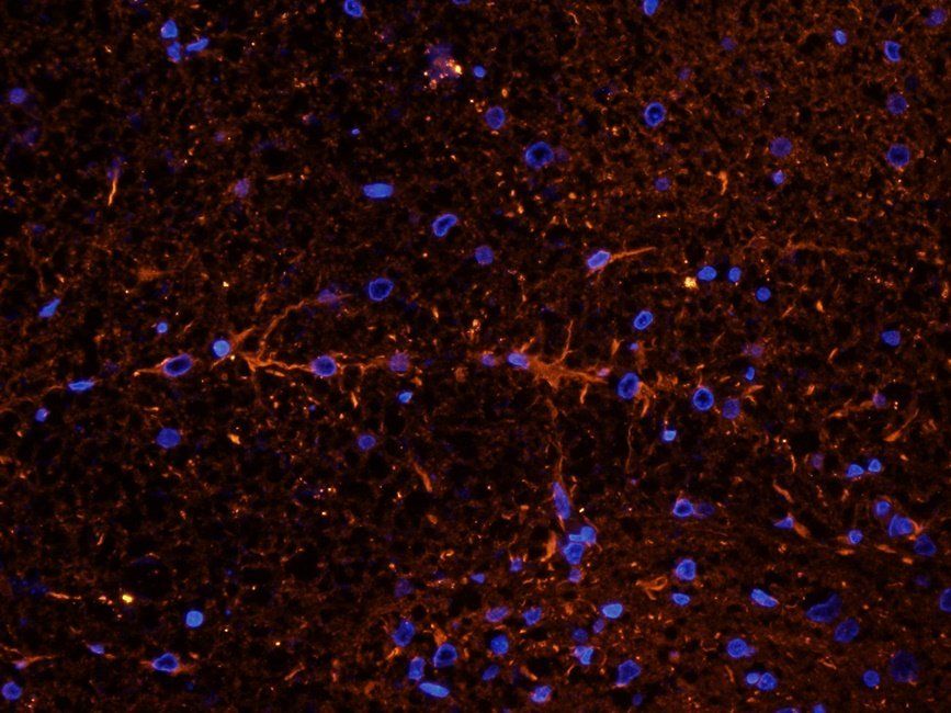
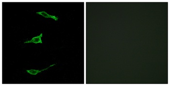
Immunofluorescence analysis of rat spinal cord tissue using FFAR1 antibody (2.5 ug/ml)
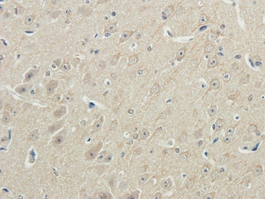
IHC-P staining of guinea pig brain tissue using anti-FFAR1 (2.5 ug/ml)
Documents Download
Datasheet
Product Information
Request a Document
Protocol Information
WB
Western Blot (IB, immunoblot)
IHC-P
Immunohistochemistry Paraffin
IF
Immunofluorescence
ICC
Immunocytochemistry
FFAR1 Rabbit Polyclonal Antibody (orb389347)

- 0.0
Based on 0 reviews
Participating in our Biorbyt product reviews program enables you to support fellow scientists by sharing your firsthand experience with our products.
Login to Submit a ReviewAvailable Sizes
Select a size below
Choose Conjugation or Carrier Free Version
Free Secondary Antibody (20 ul)0/0
Please add an antibody product to your cart first.