You have no items in your shopping cart.
Featured
Description
Research Area
Cell Biology
Images & Validation
−Item 1 of 4
| Tested Applications | ICC, IF, IHC, IP, WB |
|---|---|
| Dilution Range | WB (1:2000), IHC (1:100), ICC/IF (1:100); optimal dilutions for assays should be determined by the user. |
| Reactivity | Bovine, Canine, Guinea pig, Hamster, Human, Monkey, Mouse, Porcine, Rabbit, Rat |
| Application Notes |
Key Properties
−| Host | Mouse |
|---|---|
| Clonality | Monoclonal |
| Isotype | IgG1 |
| Clone No. | Map.ERp57 |
| Immunogen | Human recombinant ERp57 (Grp58) |
| Target | ERp57 |
| Molecular Weight | 57kDa |
| Purification | Protein G Purified |
| Conjugation | Unconjugated |
Storage & Handling
−| Storage | Maintain refrigerated at 2-8°C for up to 2 weeks. For long term storage store at -20°C in small aliquots to prevent freeze-thaw cycles. |
|---|---|
| Buffer/Preservatives | PBS pH 7.4, 50% glycerol, 0.09% sodium azide. Storage buffer changes when conjugated. |
| Concentration | 1 mg/ml |
| Expiration Date | 12 months from date of receipt. |
| Disclaimer | For research use only |
Alternative Names
−ERp60, ERp61, Grp57, Grp58, P58, PDIA3, PI PLC, 58 kDa glucose regulated protein, 58 kDa glucose-regulated protein, 58 kDa microsomal protein, Disulfide isomerase ER 60, Disulfide isomerase ER-60, Endoplasmic reticulum resident protein 57, Endoplasmic reticulum resident protein 60, ER p57, ER protein 57, ER protein 60, ERp 57, ERp57, Glucose Regulated Protein 58 Kd, GRP 57, GRP 58, GRP57, HsT17083, p58, PDIA 3, PDIA3_HUMAN, Phospholipase C alpha, Protein disulfide isomerase A3, Protein disulfide isomerase family A member 3, Protein disulfide-isomerase A3
Similar Products
−- Item 1 of 8
PDIA3 Rabbit Polyclonal Antibody [orb585694]
IHC, IP, WB
Bovine, Canine, Equine, Goat, Guinea pig, Mouse, Rabbit, Rat, Sheep, Zebrafish
Human
Rabbit
Polyclonal
Unconjugated
100 μl - Item 1 of 5
ERp57/PDIA3 Rabbit Polyclonal Antibody [orb334504]
FC, ICC, IF, IHC, IHC-Fr, WB
Human, Mouse, Rat
Rabbit
Polyclonal
Unconjugated
100 μg - Item 1 of 4
- Item 1 of 4
ERp57 rabbit pAb Antibody [orb766825]
ELISA, IF, IHC, WB
Human, Mouse, Rat
Polyclonal
Unconjugated
100 μl - Item 1 of 4
PDIA3 Antibody (Center) [orb1931335]
FC, IHC-P, WB
Bovine
Human
Rabbit
Polyclonal
Unconjugated
50 μl, 100 μl

Quality Guarantee
Explore bioreagents carefree to elevate your research. All our products are rigorously tested for performance. If a product does not perform as described on its datasheet, our scientific support team will provide expert troubleshooting, a prompt replacement, or a refund. For full details, please see our Terms & Conditions and Buying Guide. Contact us at [email protected].
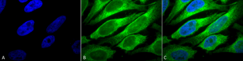
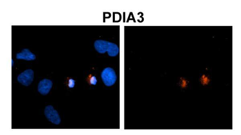
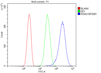
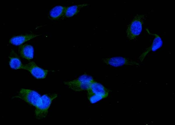
Immunocytochemistry/Immunofluorescence analysis using Mouse Anti-Erp57 (Grp58) Monoclonal Antibody, Clone Map.ERP57. Tissue: Heat Shocked cervical cancer cells (HeLa). Species: Human. Fixation: 2% Formaldehyde for 20 min at RT. Primary Antibody: Mouse Anti-Erp57 (Grp58) Monoclonal Antibody at 1:100 for 12 hours at 4°C. Secondary Antibody: FITC Goat Anti-Mouse (green) at 1:200 for 2 hours at RT. Counterstain: DAPI (blue) nuclear stain at 1:40000 for 2 hours at RT. Localization: Endoplasmic reticulum lumen. Melanosome. Magnification: 100x. (A) DAPI (blue) nuclear stain. (B) Anti-Erp57 (Grp58) Antibody. (C) Composite. Heat Shocked at 42°C for 1h.
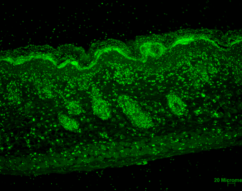
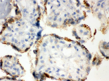
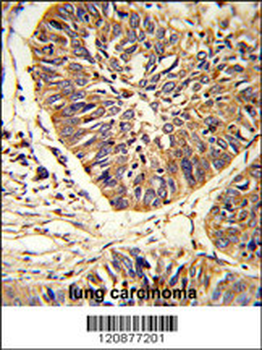
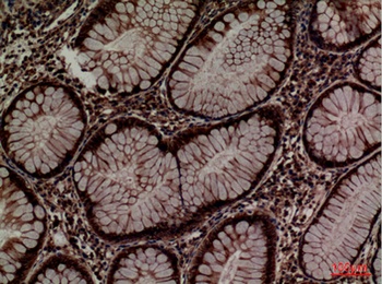
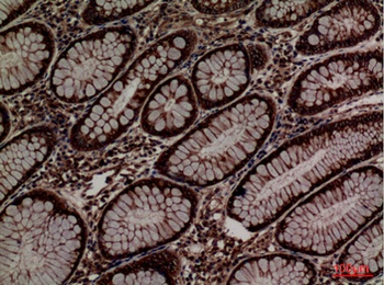
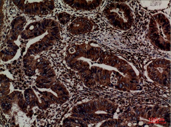
Immunohistochemistry analysis using Mouse Anti-Erp57 Monoclonal Antibody, Clone Map.ERP57. Tissue: backskin. Species: Mouse. Fixation: Bouin's Fixative and paraffin-embedded. Primary Antibody: Mouse Anti-Erp57 Monoclonal Antibody at 1:100 for 1 hour at RT. Secondary Antibody: FITC Goat Anti-Mouse (green) at 1:50 for 1 hour at RT. Localization: Epidermis and Hair Follicles.
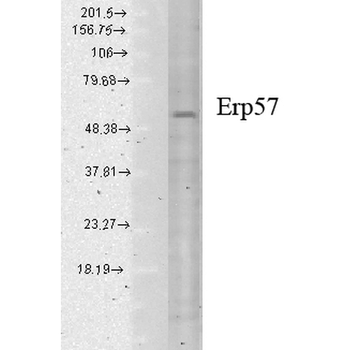
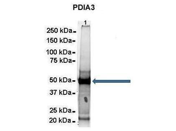
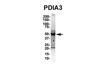
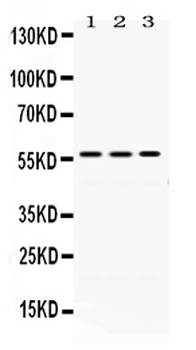
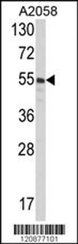
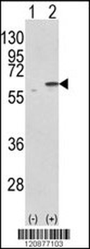
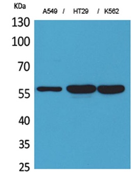
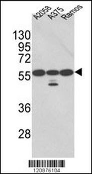
Western Blot analysis of Human cell lysates showing detection of Erp57 protein using Mouse Anti-Erp57 Monoclonal Antibody, Clone Map.ERP57. Load: 15 μg. Block: 1.5% BSA for 30 minutes at RT. Primary Antibody: Mouse Anti-Erp57 Monoclonal Antibody at 1:1000 for 2 hours at RT. Secondary Antibody: Sheep Anti-Mouse IgG: HRP for 1 hour at RT.
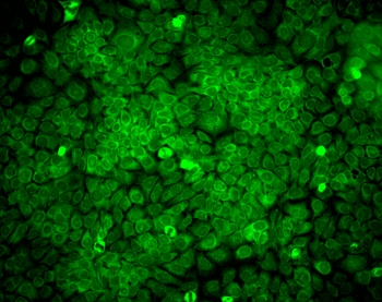
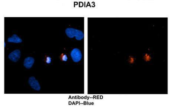
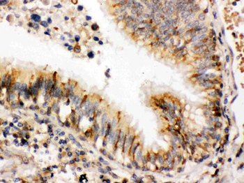
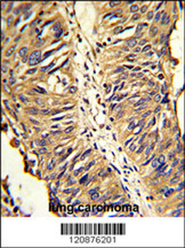
Immunocytochemistry/Immunofluorescence analysis using Mouse Anti-Erp57 Monoclonal Antibody, Clone Map.ERP57. Tissue: HaCaT cells. Species: Human. Fixation: Cold 100% methanol for 10 minutes at -20°C. Primary Antibody: Mouse Anti-Erp57 Monoclonal Antibody at 1:100 for 1 hour at RT. Secondary Antibody: FITC Goat Anti-Mouse (green) at 1:50 for 1 hour at RT. Localization: Cytoplasmic and perinuclear staining.
Quick Database Links
UniProt Details
− No UniProt data available
NCBI Gene Details
− No NCBI Gene data available
NCBI Reference Sequences
−Associated Accession Numbers
Curated reference sequences for the gene transcript and protein product| Protein | NP_005304.3 |
|---|
Documents Download
Datasheet
Product Information
Request a Document
Protocol Information
WB
Western Blot (IB, immunoblot)
IHC
Immunohistochemistry
IF
Immunofluorescence
ICC
Immunocytochemistry
IP
Immunoprecipitation
ERp57 Antibody (orb1822463)

- 0.0
Based on 0 reviews
Participating in our Biorbyt product reviews program enables you to support fellow scientists by sharing your firsthand experience with our products.
Login to Submit a ReviewAvailable Sizes
Select a size below
Free Secondary Antibody (20 ul)0/0
Please add an antibody product to your cart first.