You have no items in your shopping cart.
Featured
Description
Research Area
Stem Cell & Developmental Biology
Images & Validation
−Item 1 of 5
| Tested Applications | IF, IHC-P, WB |
|---|---|
| Dilution Range | WB: 1:1000, IF/ICC: 1:10-50, IHC-P: 1:10-50 |
| Reactivity | Human, Mouse |
Key Properties
−| Host | Rabbit |
|---|---|
| Clonality | Polyclonal |
| Isotype | Rabbit IgG |
| Immunogen | Synthetic Peptide |
| Target | EN1 |
| Molecular Weight | 40115 |
| Conjugation | Unconjugated |
Storage & Handling
−| Storage | Maintain refrigerated at 2-8°C for up to 2 weeks. For long term storage store at -20°C in small aliquots to prevent freeze-thaw cycles |
|---|---|
| Form/Appearance | Purified polyclonal antibody supplied in PBS with 0.09% (W/V) sodium azide. This antibody is purified through a protein A column, followed by peptide affinity purification. |
| Expiration Date | 12 months from date of receipt. |
| Disclaimer | For research use only |
Alternative Names
−Anti-Homeobox protein engrailed-1 antibody, anti-Hu-En-1 antibody, anti-HME1 antibody
Quality Guarantee
Explore bioreagents carefree to elevate your research. All our products are rigorously tested for performance. If a product does not perform as described on its datasheet, our scientific support team will provide expert troubleshooting, a prompt replacement, or a refund. For full details, please see our Terms & Conditions and Buying Guide. Contact us at [email protected].
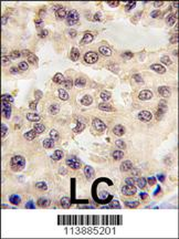
Formalin-fixed and paraffin-embedded human lung carcinoma tissue reacted with EN1 antibody (N-term), which was peroxidase-conjugated to the secondary antibody, followed by DAB staining. This data demonstrates the use of this antibody for immunohistochemistry; clinical relevance has not been evaluated.
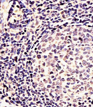
Staining EN1 in Human tonsil tissue sections by Immunohistochemistry (IHC-P - paraformaldehyde-fixed, paraffin-embedded sections). Tissue was fixed with formaldehyde and blocked with 3% BSA for 0.5 hour at room temperature; antigen retrieval was by heat mediation with a citrate buffer (pH6). Samples were incubated with primary antibody (1/25) for 1 hours at 37°C. A undiluted biotinylated goat polyvalent antibody was used as the secondary antibody.
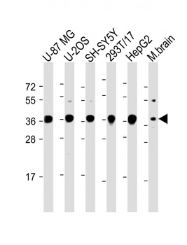
All lanes: Anti-EN1 (Engrailed 1) Antibody (N-term) at 1:2000 dilution. Lane 1: U-87 MG whole cell lysate. Lane 2: U-2OS whole cell lysate. Lane 3: SH-SY5Y whole cell lysate. Lane 4: 293T/17 whole cell lysate. Lane 5: HepG2 whole cell lysate. Lane 6: Mouse brain lysate. Lysates/proteins at 20 µg per lane. Secondary Goat Anti-Rabbit IgG, (H+L), Peroxidase conjugated at 1/10000 dilution. Predicted band size: 40 kDa. Blocking/Dilution buffer: 5% NFDM/TBST.
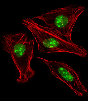
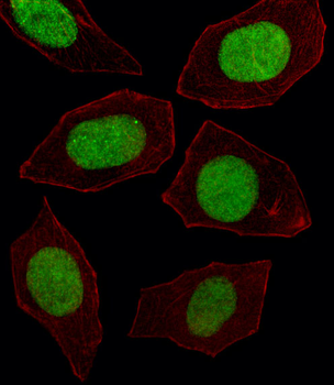
Immunofluorescent analysis of 4% paraformaldehyde-fixed, 0.1% Triton X-100 permeabilized U-2 OS (Human Sarcoma cell line) cells labeling HME1 at 1/25 dilution, followed by Alexa Fluor 488-conjugated goat anti-rabbit IgG secondary antibody at 1/400 dilution (green). Immunofluorescence image showing nucleus and nucleoli staining on U-2 OS cell line. Cytoplasmic actin is detected with Alexa Fluor 555 conjugated with Phalloidin at 1/100 dilution (red).
Fluorescent image of U251 cell stained with EN1 Antibody (N-term). U251 cells were fixed with 4% PFA (20 min), permeabilized with Triton X-100 (0.1%, 10 min), then incubated with EN1 primary antibody (1:25, 1 h at 37°C). For secondary antibody, Alexa Fluor 488 conjugated donkey anti-rabbit antibody (green) was used (1:400, 50 min at 37°C). Cytoplasmic actin was counterstained with Alexa Fluor 555 (red) conjugated Phalloidin (7 units/ml, 1 h at 37°C). EN1 immunoreactivity is localized to Nucleus significantly.
Quick Database Links
UniProt Details
− No UniProt data available
NCBI Reference Sequences
−Associated Accession Numbers
Curated reference sequences for the gene transcript and protein product| Protein | NP_001417.3 |
|---|
Documents Download
Datasheet
Product Information
Request a Document
Protocol Information
WB
Western Blot (IB, immunoblot)
IHC-P
Immunohistochemistry Paraffin
IF
Immunofluorescence
RJ Xu Epigenetic and Transcriptional Regulations in Pancreatic Ductal Adenocarcinoma search.proquest.com, (2024)
EN1 (Engrailed 1) Antibody (N-term) (orb37720)

- 0.0
Based on 0 reviews
Participating in our Biorbyt product reviews program enables you to support fellow scientists by sharing your firsthand experience with our products.
Login to Submit a ReviewAvailable Sizes
Select a size below
Choose Conjugation or Carrier Free Version
Free Secondary Antibody (20 ul)0/0
Please add an antibody product to your cart first.