You have no items in your shopping cart.
Featured
KO/KD
Validated
Validated
Description
Research Area
Cell Biology
Images & Validation
−Item 1 of 5
| Tested Applications | ELISA, IF, IHC-P, KO/KD Validated, WB |
|---|---|
| Reactivity | Human, Mouse, Rat |
Key Properties
−| Antibody Type | Primary Antibody |
|---|---|
| Host | Rabbit |
| Clonality | Polyclonal |
| Isotype | IgG |
| Immunogen | Anti-DRAM antibody (orb1239379) was raised against a peptide corresponding to 16 amino acids near the carboxy terminus of human DRAM. The immunogen is located within amino acids 170-220 of DRAM. |
| Target | DRAM1 |
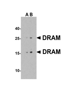
| Molecular Weight | Predicted: 14kD, 26kDObserved: 14 kD, 26kD |
| Purification | DRAM Antibody is affinity chromatography purified via peptide column. |
| Conjugation | Unconjugated |
Storage & Handling
−| Storage | Maintain refrigerated at 2-8°C for up to 2 weeks. For long term storage store at -20°C in small aliquots to prevent freeze-thaw cycles. |
|---|---|
| Form/Appearance | Liquid |
| Buffer/Preservatives | DRAM Antibody is supplied in PBS containing 0.02% sodium azide. |
| Concentration | 1 mg/mL |
| Expiration Date | 12 months from date of receipt. |
| Disclaimer | For research use only |
Alternative Names
−DRAM Antibody: DRAM, DRAM, DNA damage-regulated autophagy modulator protein 1, Damage-regulated autophagy modulator
Similar Products
−- Item 1 of 6
DRAM1 Antibody [orb1239371]
ELISA, IF, IHC-P, WB
Human, Mouse, Rat
Rabbit
Polyclonal
Unconjugated
0.02 mg, 0.1 mg - Item 1 of 2
DRAM1/DRAM Antibody [orb1537868]
ELISA, IF, IHC, IHC-P, WB
Human, Mouse, Rat
Rabbit
Polyclonal
Unconjugated
0.05 mg - Item 1 of 2
- Item 1 of 2
DRAM Rabbit Polyclonal Antibody [orb183254]
ICC, IF, IHC-Fr, IHC-P
Canine, Equine, Mouse, Porcine, Rabbit, Sheep
Human, Rat
Rabbit
Polyclonal
Unconjugated
200 μl, 50 μl, 100 μl - Item 1 of 1
DRAM1 rabbit pAb Antibody [orb774311]
ELISA, WB
Human, Mouse, Rat
Polyclonal
Unconjugated
50 μl, 100 μl


Quality Guarantee
Explore bioreagents carefree to elevate your research. All our products are rigorously tested for performance. If a product does not perform as described on its datasheet, our scientific support team will provide expert troubleshooting, a prompt replacement, or a refund. For full details, please see our Terms & Conditions and Buying Guide. Contact us at [email protected].

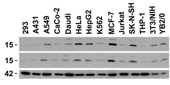
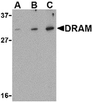
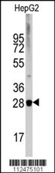
Independent Antibody Validation (IAV) via Protein Expression Profile in Cell Lines. Loading: 15 µg of lysates per lane. Antibodies: DRAM orb1239379 (0.5 µg/mL), DRAM orb1239371 (2 µg/mL), beta-actin (1 µg/mL) and GAPDH (0.02 µg/mL), 1h incubation at RT in 5% NFDM/TBST. Secondary: Goat anti-rabbit IgG HRP conjugate at 1:10000 dilution.
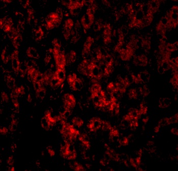
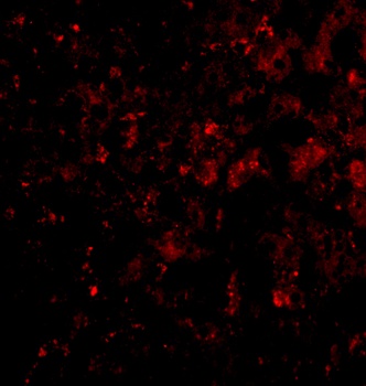
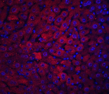
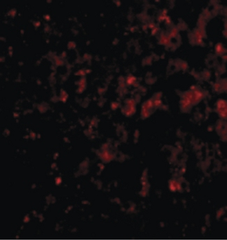
Immunofluorescence Validation of DRAM in Human Liver Tissue. Immunofluorescent analysis of 4% paraformaldehyde-fixed human liver tissue labeling DRAM with orb1239379 at 20 µg/mL, followed by goat anti-rabbit IgG secondary antibody at 1/500 dilution (red).
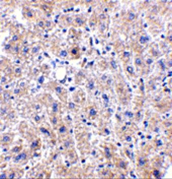
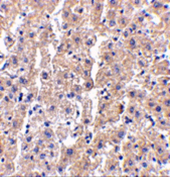
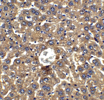
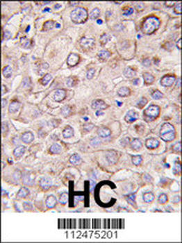
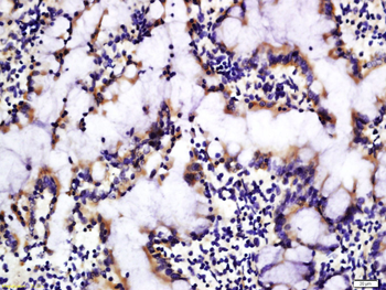
Immunohistochemistry Validation of DRAM in Human Liver Tissue. Immunohistochemical analysis of paraffin-embedded human liver tissue using anti-DRAM antibody (orb1239379) at 2.5 µg/ml. Tissue was fixed with formaldehyde and blocked with 10% serum for 1 h at RT; antigen retrieval was by heat mediation with a citrate buffer (pH6). Samples were incubated with primary antibody overnight at 4°C. A goat anti-rabbit IgG H&L (HRP) at 1/250 was used as secondary. Counter stained with Hematoxylin.
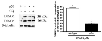
Induced Expression of DRAM by CQ in Mouse Neural Precursor Cells (NPCs) (Walls et al., 2010). WT and p53-deficient NPC cells were treated with or without 25µM CQ for 24hr. CQ treatment caused the increased expression level in both DRAM dimer (32kD) and monomer (16kD) compared to the untreated controls. WB results show DRAM induction was p53-dependent.
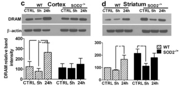
Regulated Expression Validation of DRAM in Heterozygous SOD2 KO Mice (Mehta et al., 2011). DRAM expression level detected by anti-DRAM antibodies (orb1239379) decreased in striatum of SOD -/+ KO mice (fig. d) as compared to WT mice.
Documents Download
Datasheet
Product Information
Request a Document
Protocol Information
WB
Western Blot (IB, immunoblot)
IHC-P
Immunohistochemistry Paraffin
IF
Immunofluorescence
ELISA
Enzyme-linked Immunosorbent Assay (EIA)
DRAM1 Antibody (orb1239379)

- 0.0
Based on 0 reviews
Participating in our Biorbyt product reviews program enables you to support fellow scientists by sharing your firsthand experience with our products.
Login to Submit a ReviewAvailable Sizes
Select a size below
Free Secondary Antibody (20 ul)0/0
Please add an antibody product to your cart first.