You have no items in your shopping cart.
Description
Research Area
Signal Transduction
Images & Validation
−Item 1 of 4
| Tested Applications | IF, IHC-P, IP, WB |
|---|---|
| Dilution Range | IF - 1:10-50, IP - 1:500-1000, WB - 1:1000, IHC-P - 1:10-50 |
| Reactivity | Human, Mouse |
Key Properties
−| Host | Rabbit |
|---|---|
| Clonality | Polyclonal |
| Isotype | Rabbit IgG |
| Immunogen | This DCK antibody is generated from rabbits immunized with a KLH conjugated synthetic peptide between 171-200 amino acids from the C-terminal region of human DCK. Antigen Region: 171-200 aa. |
| Target | DCK |
| Molecular Weight | 30519 Da |
| Conjugation | Unconjugated |
Storage & Handling
−| Storage | Maintain refrigerated at 2-8°C for up to 2 weeks. For long term storage store at -20°C in small aliquots to prevent freeze-thaw cycles |
|---|---|
| Form/Appearance | Purified polyclonal antibody supplied in PBS with 0.09% (W/V) sodium azide. This antibody is prepared by Saturated Ammonium Sulfate (SAS) precipitation followed by dialysis against PBS. |
| Expiration Date | 12 months from date of receipt. |
| Disclaimer | For research use only |
Alternative Names
−Deoxycytidine kinase, dCK, DCK
Quality Guarantee
Explore bioreagents carefree to elevate your research. All our products are rigorously tested for performance. If a product does not perform as described on its datasheet, our scientific support team will provide expert troubleshooting, a prompt replacement, or a refund. For full details, please see our Terms & Conditions and Buying Guide. Contact us at [email protected].
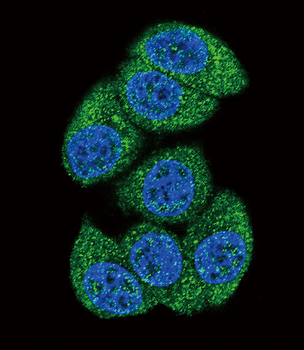
Confocal immunofluorescent analysis of DCK Antibody (C-term) with Hela cell followed by Alexa Fluor 488-conjugated goat anti-rabbit lgG (green). DAPI was used to stain the cell nuclear (blue).
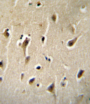
DCK Antibody (C-term) immunohistochemistry analysis in formalin fixed and paraffin embedded human brain tissue followed by peroxidase conjugation of the secondary antibody and DAB staining. This data demonstrates the use of DCK Antibody (C-term) for immunohistochemistry. Clinical relevance has not been evaluated.
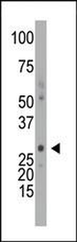
The anti-DCK Pab is used in Western blot to detect DCK in mouse intestine tissue lysate.
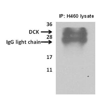
Deoxycytidine kinase (DCK) immunoprecipitated from H460cells with 7.5ug (microgram) of the dCK antibody using the Pierce classic mammalian IP kit reagent as described as manufacturer instructions (lane 1, 3) and Current Protocols in Cell Biology, 1998, 7.2.1-7.2.21. Proteins separated on a 12% SDS gel, transferred to a PVDF membrane and probed with 1: 700 dilution of DCK antibody. Bands were detected using enhanced chemiluminescence (SuperSignal West Pico Chemiluminescent Substrate Kit). No specific reagents were employed to remove IgG from immunoprecipitated sample.
Quick Database Links
UniProt Details
− No UniProt data available
NCBI Reference Sequences
−Associated Accession Numbers
Curated reference sequences for the gene transcript and protein product| Protein | NP_000779.1 |
|---|
Documents Download
Datasheet
Product Information
Request a Document
Protocol Information
WB
Western Blot (IB, immunoblot)
IHC-P
Immunohistochemistry Paraffin
IF
Immunofluorescence
IP
Immunoprecipitation
DCK Antibody (C-term) (orb1929569)

- 0.0
Based on 0 reviews
Participating in our Biorbyt product reviews program enables you to support fellow scientists by sharing your firsthand experience with our products.
Login to Submit a ReviewAvailable Sizes
Select a size below
Free Secondary Antibody (20 ul)0/0
Please add an antibody product to your cart first.