You have no items in your shopping cart.
Featured
Description
Research Area
Cell Cycle, Cyclin b Family, Cyclins
Images & Validation
−Item 1 of 9
| Tested Applications | FC, ICC, IF, IHC-Fr, IHC-P, WB |
|---|---|
| Dilution Range | WB=1:500-2000, IHC-P=1:100-500, IHC-F=1:100-500, ICC/IF=1:100-500, IF=1:100-500, Flow-Cyt=1μg/Test |
| Reactivity | Human, Mouse, Rat |
| Predicted Reactivity | Bovine |
Related Conjugates & Formulations
−Key Properties
−| Antibody Type | Primary Antibody |
|---|---|
| Host | Rabbit |
| Clonality | Polyclonal |
| Isotype | IgG |
| Immunogen | KLH conjugated synthetic peptide derived from human Cyclin B1 (271-433/433aa) |
| Target | CCNB1 |
| Molecular Weight | 50 kDa |
| Purification | Affinity purified by Protein A |
| Conjugation | Unconjugated |
Storage & Handling
−| Storage | Maintain refrigerated at 2-8°C for up to 2 weeks. For long term storage store at -20°C in small aliquots to prevent freeze-thaw cycles. |
|---|---|
| Form/Appearance | Liquid |
| Buffer/Preservatives | 0.01M TBS (pH7.4) with 1% rAlbumin, 0.02% Proclin300 and 50% Glycerol. |
| Concentration | 1mg/ml |
| Expiration Date | 12 months from date of receipt. |
| Disclaimer | For research use only |
Alternative Names
−CCNB; Ccnb1-rs1; Ccnb1-rs13; CycB1; Cycb-4; Cycb-5; Cycb1-rs1; CCNB1_HUMAN; CCNB1; CCNB1_MOUSE; Ccn-2; Cycb; CCNB1_RAT; cyclin B1; G2/mitotic-specific cyclin B1
Similar Products
−- Item 1 of 10
Cyclin B1/CCNB1 Rabbit Polyclonal Antibody [orb654315]
ELISA, FC, ICC, IF, IHC, WB
Human, Mouse, Rat
Rabbit
Polyclonal
Unconjugated
100 μg - Item 1 of 4
Cyclin B1 (phospho Ser126) rabbit pAb Antibody [orb764169]
ELISA, IF, IHC, WB
Human, Mouse, Rat
Polyclonal
Unconjugated
50 μl, 100 μl - Item 1 of 4
Phospho-Cyclin B1 (Ser126) Rabbit Polyclonal Antibody [orb5928]
ICC, IF, IHC-Fr, IHC-P
Human
Rabbit
Polyclonal
Unconjugated
50 μl, 100 μl, 200 μl - Item 1 of 4
CCNB1 Rabbit Polyclonal Antibody [orb626160]
ELISA, IF, IHC, IP, WB
Human, Mouse, Rat
Rabbit
Polyclonal
Unconjugated
50 μg, 100 μg - Item 1 of 4
Cyclin B1 Rabbit Polyclonal Antibody [orb1294340]
IF, IHC, WB
Human
Rabbit
Polyclonal
Unconjugated
25 μl, 100 μl


Quality Guarantee
Explore bioreagents carefree to elevate your research. All our products are rigorously tested for performance. If a product does not perform as described on its datasheet, our scientific support team will provide expert troubleshooting, a prompt replacement, or a refund. For full details, please see our Terms & Conditions and Buying Guide. Contact us at [email protected].
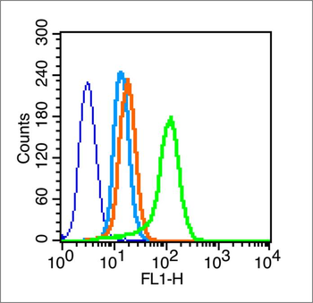
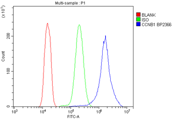
Blank control (blue line): A549 (blue). Primary Antibody (green line): Rabbit Anti-Cyclin B1 antibody (orb10494). Dilution: 1 µg/10^6 cells, Isotype Control Antibody (orange line): Rabbit IgG. Secondary Antibody (white blue line): F (ab')2 fragment goat anti-rabbit IgG-FITC. Dilution: 1 µg/Test. Protocol, The cells were fixed with 2% paraformaldehyde (10 min) and then permeabilized with 0.1% PBS-Tween for 20 min at room temperature. Cells stained with Primary Antibody for 30 min at room temperature. The cells were then incubated in 1 X PBS/2% BSA/10% goat serum to block non-specific protein-protein interactions followed by the antibody for 15 min at room temperature. The secondary antibody used for 40 min at room temperature. Acquisition of 20000 events was performed.
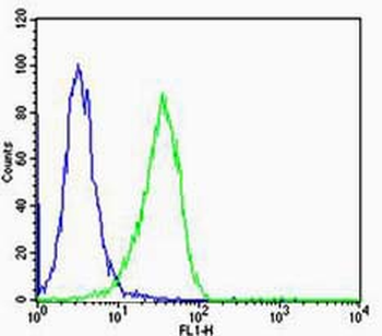
Cell: Hela, Concentration: 1:100, Host/Isotype: Rabbit/IgG, Flow cytometric analysis of primary antibody (Cat#: orb10494) on Hela (green) compared with isotype control in the absence of primary antibody (blue) followed by Alexa Fluor 488-conjugated goat anti-rabbit IgG (H+L) secondary antibody.
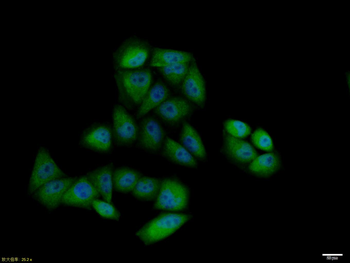
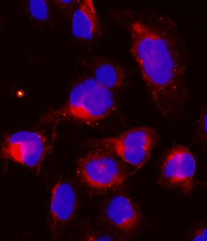
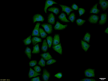
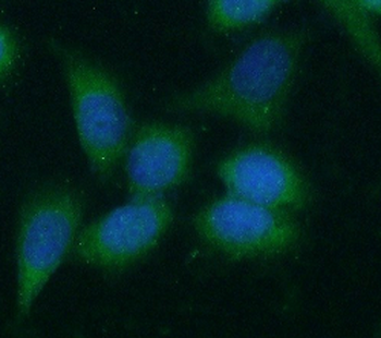
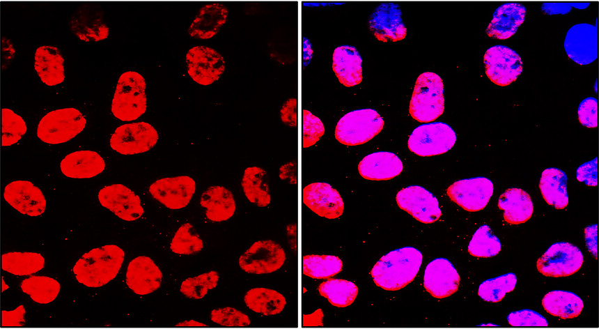
Hela cell, 4% Paraformaldehyde-fixed, Triton X-100 at room temperature for 20 min, Blocking buffer (normal goat serum) at 37°C for 20 min, Antibody incubation with (Cyclin B1) polyclonal Antibody, Unconjugated (orb10494) 1:100, 90 minutes at 37°C, followed by a conjugated Goat Anti-Rabbit IgG antibody at 37°C for 90 minutes, DAPI (blue) was used to stain the cell nuclei.
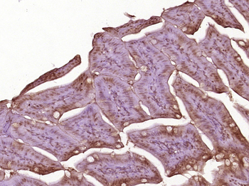
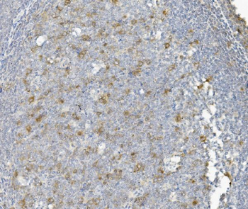
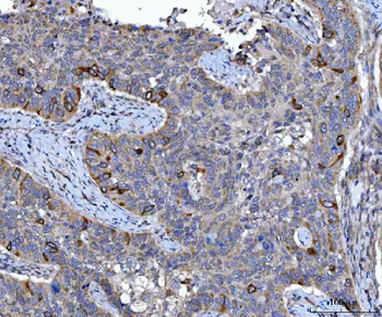
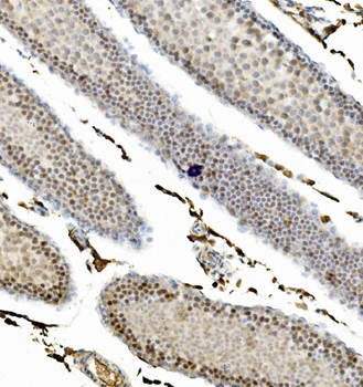
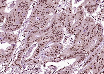
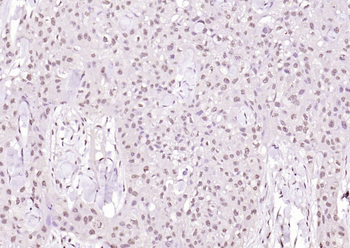
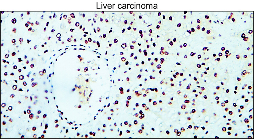
Paraformaldehyde-fixed, paraffin embedded (Mouse small intestine), Antigen retrieval by boiling in sodium citrate buffer (pH6.0) for 15 min, Block endogenous peroxidase by 3% hydrogen peroxide for 20 minutes, Blocking buffer (normal goat serum) at 37°C for 30 min, Antibody incubation with (Cyclin B1) Polyclonal Antibody, Unconjugated (orb10494) at 1:400 overnight at 4°C, followed by operating according to SP Kit (Rabbit) instructionsand DAB staining.
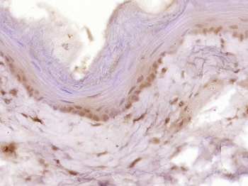
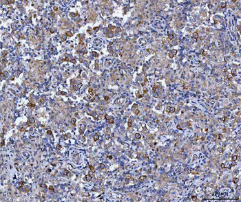
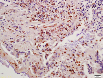
Paraformaldehyde-fixed, paraffin embedded (Rat esophageal), Antigen retrieval by boiling in sodium citrate buffer (pH6.0) for 15 min, Block endogenous peroxidase by 3% hydrogen peroxide for 20 minutes, Blocking buffer (normal goat serum) at 37°C for 30 min, Antibody incubation with (Cyclin B1) Polyclonal Antibody, Unconjugated (orb10494) at 1:400 overnight at 4°C, followed by operating according to SP Kit (Rabbit) instructionsand DAB staining.
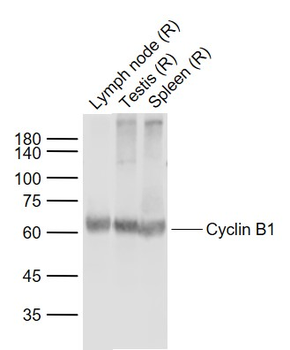
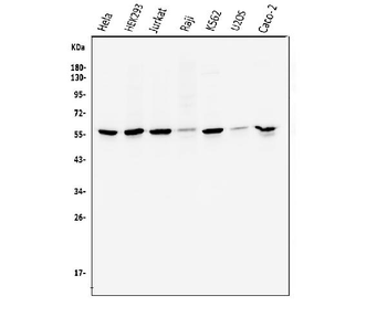
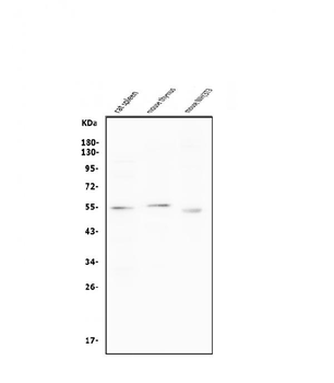
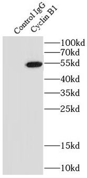
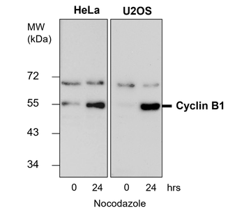
Sample: Lane 1: Lymph node (Rat) Lysate at 40 ug, Lane 2: Testis (Rat) Lysate at 40 ug, Lane 3: Spleen (Rat) Lysate at 40 ug, Primary: Anti-Cyclin B1 (orb10494) at 1/1000 dilution, Secondary: IRDye800CW Goat Anti-Rabbit IgG at 1/20000 dilution, Predicted band size: 55-60 kD, Observed band size: 60 kD.
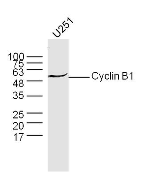
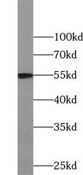
Sample: U251 Cell (Human) Lysate at 30 ug, Primary: Anti-Cyclin B1 (orb10494) at 1/300 dilution, Secondary: IRDye800CW Goat Anti-Rabbit IgG at 1/20000 dilution, Predicted band size: 48 kD, Observed band size: 50 kD.
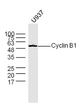
Sample: U937 Cell (Human) Lysate at 30 ug, Primary: Anti-Cyclin B1 (orb10494) at 1/300 dilution, Secondary: IRDye800CW Goat Anti-Rabbit IgG at 1/20000 dilution, Predicted band size: 48 kD, Observed band size: 50 kD.
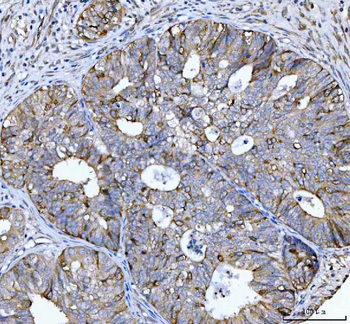
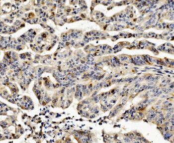
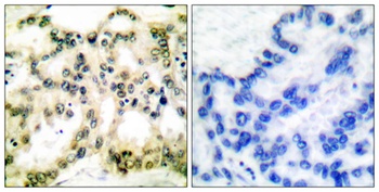
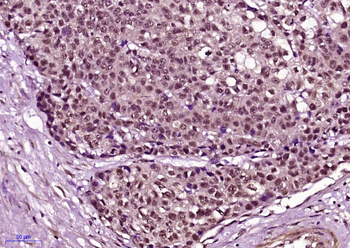
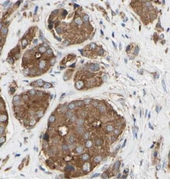
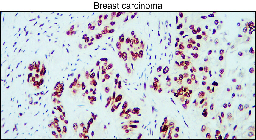
Tissue/Cell: human colon carcinoma, 4% Paraformaldehyde-fixed and paraffin-embedded, Antigen retrieval: citrate buffer (0.01M, pH6.0), Boiling bathing for 15 min, Block endogenous peroxidase by 3% Hydrogen peroxide for 30 min, Blocking buffer (normal goat serum) at 37°C for 20 min, Incubation: Anti-Cyclin B1 Polyclonal Antibody, Unconjugated (orb10494) 1:200, overnight at 4°C, followed by conjugation to the secondary antibody and DAB staining.
Quick Database Links
Gene Symbol
CCNB1
UniProt
UniProt Details
− No UniProt data available
Documents Download
Datasheet
Product Information
Request a Document
Protocol Information
WB
Western Blot (IB, immunoblot)
IHC-P
Immunohistochemistry Paraffin
IHC-Fr
Immunohistochemistry Frozen
FC
Flow Cytometry
IF
Immunofluorescence
ICC
Immunocytochemistry
Wang, Qi et al. Methamphetamine induces hepatotoxicity via inhibiting cell division, arresting cell cycle and activating apoptosis: In vivo and in vitro studies Food Chem Toxicol, 105, 61-72 (2017)
Cyclin B1 Rabbit Polyclonal Antibody (orb10494)

- 0.0
Based on 0 reviews
Participating in our Biorbyt product reviews program enables you to support fellow scientists by sharing your firsthand experience with our products.
Login to Submit a ReviewAvailable Sizes
Select a size below
Free Secondary Antibody (20 ul)0/0
Please add an antibody product to your cart first.