You have no items in your shopping cart.
Description
Research Area
Stem Cell & Developmental Biology
Images & Validation
−Item 1 of 6
| Tested Applications | IF, IHC-P, WB |
|---|---|
| Dilution Range | WB - 1:2000, IF - 1:25, IHC-P - 1:25 |
| Reactivity | Human |
Key Properties
−| Host | Rabbit |
|---|---|
| Clonality | Polyclonal |
| Isotype | Rabbit IgG |
| Immunogen | This CUX1 antibody is generated from rabbits immunized with a KLH conjugated synthetic peptide between 1347-1374 amino acids of human CUX1. Antigen Region: 1347-1374 aa. |
| Target | CUX1 (HGNC:2557) |
| Molecular Weight | 164187 Da |
| Conjugation | Unconjugated |
Storage & Handling
−| Storage | Maintain refrigerated at 2-8°C for up to 2 weeks. For long term storage store at -20°C in small aliquots to prevent freeze-thaw cycles |
|---|---|
| Form/Appearance | Purified polyclonal antibody supplied in PBS with 0.09% (W/V) sodium azide. This antibody is prepared by Saturated Ammonium Sulfate (SAS) precipitation followed by dialysis against PBS. |
| Expiration Date | 12 months from date of receipt. |
| Disclaimer | For research use only |
Alternative Names
−Homeobox protein cut-like 1, CCAAT displacement protein, CDP, Homeobox protein cux-1, CUX1, CUTL1
Similar Products
−- Item 1 of 1

Quality Guarantee
Explore bioreagents carefree to elevate your research. All our products are rigorously tested for performance. If a product does not perform as described on its datasheet, our scientific support team will provide expert troubleshooting, a prompt replacement, or a refund. For full details, please see our Terms & Conditions and Buying Guide. Contact us at [email protected].
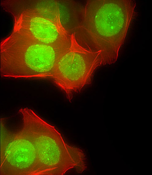
Immunofluorescent analysis of 4% paraformaldehyde-fixed, 0.1% Triton X-100 permeabilized MCF-7 (human breast cancer cell line) cells labeling CUX1 at 1/25 dilution, followed by Dylight 488-conjugated goat anti-rabbit IgG secondary antibody at 1/200 dilution (green). Immunofluorescence image showing nucleus and weak cytoplasm staining on MCF-7 cell line. Cytoplasmic actin is detected with Dylight 554 Phalloidin at 1/100 dilution (red).
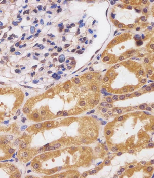
Staining CUX1 in human kidney tissue sections by Immunohistochemistry (IHC-P - paraformaldehyde-fixed, paraffin-embedded sections). Tissue was fixed with formaldehyde and blocked with 3% BSA for 0.5 hour at room temperature; antigen retrieval was by heat mediation with a citrate buffer (pH6). Samples were incubated with primary antibody (1/25) for 1 hours at 37°C. A undiluted biotinylated goat polyvalent antibody was used as the secondary antibody.
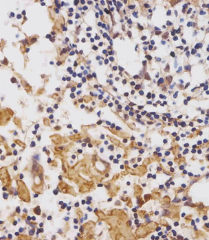
Staining CUX1 in human lymph node tissue sections by Immunohistochemistry (IHC-P - paraformaldehyde-fixed, paraffin-embedded sections). Tissue was fixed with formaldehyde and blocked with 3% BSA for 0.5 hour at room temperature; antigen retrieval was by heat mediation with a citrate buffer (pH6). Samples were incubated with primary antibody (1/25) for 1 hours at 37°C. A undiluted biotinylated goat polyvalent antibody was used as the secondary antibody.
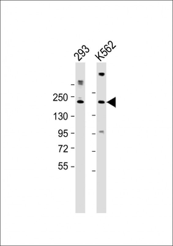
All lanes: Anti-CUX1 Antibody (C-term) at 1:2000 dilution. Lane 1: 293 whole cell lysate. Lane 2: K562 whole cell lysate. Lysates/proteins at 20 µg per lane. Secondary Goat Anti-Rabbit IgG, (H+L), Peroxidase conjugated at 1/10000 dilution. Predicted band size: 164 kDa. Blocking/Dilution buffer: 5% NFDM/TBST.
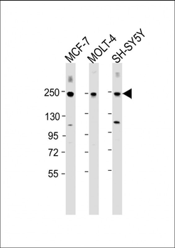
All lanes: Anti-CUX1 Antibody (C-term) at 1:2000 dilution. Lane 1: MCF-7 whole cell lysate. Lane 2: MOLT-4 whole cell lysate. Lane 3: SH-SY5Y whole cell lysate. Lysates/proteins at 20 µg per lane. Secondary Goat Anti-Rabbit IgG, (H+L), Peroxidase conjugated at 1/10000 dilution. Predicted band size: 164 kDa. Blocking/Dilution buffer: 5% NFDM/TBST.
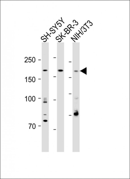
All lanes: Anti-CUX1 Antibody (C-term) at 1:1000 dilution. Lane 1: SH-SY5Y whole cell lysate. Lane 2: SK-BR-3 whole cell lysate. Lane 3: NIH/3T3 whole cell lysate. Lysates/proteins at 20 µg per lane. Secondary: Goat Anti-Rabbit IgG, (H+L), Peroxidase conjugated at 1/15000 dilution. Observed band size: 200 KDa. Blocking/Dilution buffer: 5% NFDM/TBST.
Quick Database Links
Gene Symbol
CUX1 (HGNC:2557)
UniProt
RefSeq (Protein):NP_001189472.1, NP_852477.1, NP_001189474.1, NP_853530.2, NP_001189473.1
UniProt Details
− No UniProt data available
NCBI Reference Sequences
−Associated Accession Numbers
Curated reference sequences for the gene transcript and protein productDocuments Download
Datasheet
Product Information
Request a Document
Protocol Information
WB
Western Blot (IB, immunoblot)
IHC-P
Immunohistochemistry Paraffin
IF
Immunofluorescence
CUX1 Antibody (C-term) (orb1930297)

- 0.0
Based on 0 reviews
Participating in our Biorbyt product reviews program enables you to support fellow scientists by sharing your firsthand experience with our products.
Login to Submit a ReviewAvailable Sizes
Select a size below
Free Secondary Antibody (20 ul)0/0
Please add an antibody product to your cart first.