You have no items in your shopping cart.
Description
Research Area
Cardiovascular Research
Images & Validation
−Item 1 of 6
| Tested Applications | ELISA, IHC, IP, WB |
|---|---|
| Dilution Range | ELISA: 1:20,000 - 1:100,000, IHC: 1:1,000 - 1:5,000, IP: 1:100, WB: 1:2,000 - 1:10,000 |
| Reactivity | Bovine, Human |
| Application Notes |
Key Properties
−| Antibody Type | Primary Antibody |
|---|---|
| Host | Rabbit |
| Clonality | Polyclonal |
| Isotype | IgG |
| Immunogen | Anti-Collagen Type IV was produced by repeated immunizations with Collagen Type IV from human and bovine placenta. |
| Target | COL4A1-COL4A6 |
| Purity | Anti-Collagen Type IV has been prepared by immunoaffinity chromatography using immobilized antigens followed by extensive cross-adsorption against other collagens, human serum proteins and non-collagen extracellular matrix proteins to remove any unwanted specificities. Some class-specific anti-collagens may be specific for three-dimensional epitopes which may result in diminished reactivity with denatured collagen or formalin-fixed, paraffin embedded tissues. This antibody reacts with most mammalian Type IV collagens and has negligible cross-reactivity with Type I, II, III, V and VI collagens. Non-specific cross-reaction of anti-collagen antibodies with other human serum proteins or non-collagen extracellular matrix proteins is negligible. |
| Conjugation | Biotin |
Storage & Handling
−| Storage | Store vial at 4° C prior to restoration. Restore with 0.1 mL of deionized water (or equivalent). For extended storage aliquot contents and freeze at -20° C or below. Avoid cycles of freezing and thawing. Centrifuge product if not completely clear after standing at room temperature. This product is stable for several weeks at 4° C as an undiluted liquid. Dilute only prior to immediate use. Expiration date is one (1) year from date of restoration. |
|---|---|
| Form/Appearance | Lyophilized |
| Buffer/Preservatives | Preservative: 0.01% (w/v) Sodium Azide. Stabilizer: 10 mg/mL Bovine Serum Albumin (rAlbumin) - Immunoglobulin and Protease free; Buffer: 0.02 M Potassium Phosphate, 0.15 M Sodium Chloride, pH 7.2 |
| Concentration | 1.0 mg/mL |
| Expiration Date | 12 months from date of receipt. |
| Disclaimer | For research use only |
Alternative Names
−rabbit anti-Collagen Type IV antibody biotin conjugation, biotin conjugated rabbit anti-Collagen Type IV antibody, Arresten antibody, Canstatin antibody, Collagen Of Basement Membrane Alpha 1 Chain antibody, Collagen alpha-1 (IV) chain, COL4A1
Quality Guarantee
Explore bioreagents carefree to elevate your research. All our products are rigorously tested for performance. If a product does not perform as described on its datasheet, our scientific support team will provide expert troubleshooting, a prompt replacement, or a refund. For full details, please see our Terms & Conditions and Buying Guide. Contact us at [email protected].
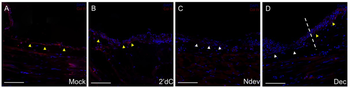
Immunohistochemistry of Anti-Collagen Type IV Antibody Biotin Conjugated. Inhibition of Collagen IV expression in decitabine-treated wound epithelia. Immunohistochemistry staining for Collagen IV, a marker for the basal lamina, in wounds six days post-surgery. Wounds were either untreated (A, mock); received an implanted bead containing 2'deoxycytidine without a deviated nerve (B, 2'dC); received a surgically deviated nerve to induce formation of an ectopic blastema (C, NDev); or received an implanted bead containing decitabine without a deviated nerve (D, Dec). White arrowheads indicate areas within the wound that are negative for ColIV staining; yellow arrowheads indicate areas that are positive for Col IV staining. Dotted line in (D) indicates the transition between the uninjured skin (right) and the wound (left). Scale bars = 200 microns.
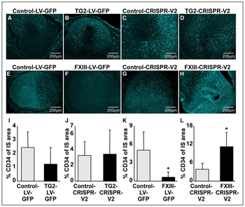
Immunohistochemistry of Anti-Collagen Type IV Antibody Biotin Conjugated. Microvascular evaluation of the implantation site (IS) with transgenic embryos. Immunostaining for CD34 (cluster of differentiation 34) in ISs of genetically modified trophoblast cells (TC) expressing the control vector (LV-GFP [lentivirusgreen fluorescent protein], A and E; CRISPR-V2, C and G, both 4 dams, 10 ISs), overexpressing tissue TG (TG2) (TG2-LV-GFP; B, 4 dams, 12 ISs), depleted from TG2 (tissue TG; D, 3 dams, 10 ISs), overexpressing FXIII (factor XIII; F, 5 dams, 19 ISs) or depleted from FXIII (H, 4 dams, 12 ISs). CD34 was visualized by cyan fluorescence channel after labeled with Cy5-avidin. Similar exposure time was used for each section and its appropriate control. Quantitative analysis of% CD34 staining in relation to IS area of genetically modified TC overexpressing TG2 (I), depleted from TG2 (J), overexpressing FXIII (K) or depleted from FXIII (L). Image scale bars are 200 µm. Fibrinogen was clearly detected in the anti-mesometrial pole and adjacent to control embryonic TC (Figure 5A, upper part). Increased fibrinogen deposition was detected in IS of FXIII-overexpressing TC, particularly in the IS circumference, while fibrinogen was diminished at the embryonic vicinity (Figure 5B, upper part), as compared with control. CIV localization on the control IS (Figure 5A, lower part) was confined to the anti-mesometrial pole and around the primary decidual zone, while a substantial wider partition was displayed on the FXIII overexpressed TC (Figure 5B, lower part). Inversely to the IS with FXIII overexpressed TC, fibrinogen and Collagen Type IV were hardly detected in IS with FXIII-depleted TC (Figure 5D) relative to control (Figure 5C).
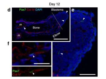
Immunohistochemistry of Anti-Collagen Type IV Antibody Biotin Conjugated. SMFC tracking in larval newt limb regeneration. (d–f) Pax7 immunolabelling of regenerating limbs on day 12 (n ¼ 3) after amputation. (d) On day 12, a few Pax7 þ nuclei (arrowheads) were detected in blastema cells and in satellite cells along the muscle fibers. Col IV, collagen type IV immunoreactivity. DAPI (4, 6-diamidino-2-phenylindole), nuclei. Scale bar, 300 mm. The Pax7 þ nuclei pointed by arrowheads were enlarged in e and f, respectively. Scale bars, 100 mm.
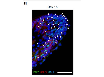
Immunohistochemistry of Anti-Collagen Type IV Antibody Biotin Conjugated. SMFC tracking in larval newt limb regeneration. (g) On day 15 when the regenerating part of the limb grew more distally, the number of Pax7 þ nuclei (arrowheads) in the blastema was dramatically increased. Scale bar, 100 mm.
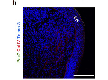
Immunohistochemistry of Anti-Collagen Type IV Antibody Biotin Conjugated. SMFC tracking in metamorphosed newt limb regeneration. Merged fluorescence image. Pax7; Col IV, collagen type IV immunoreactivity; To-pro-3: nuclei. (h) Enlargement of a region in the blastema. Scale bars, 250 mm.
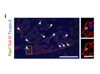
Immunohistochemistry of Anti-Collagen Type IV Antibody Biotin Conjugated. SMFC tracking in metamorphosed newt limb regeneration. Merged fluorescence image. Pax7; Col IV, collagen type IV immunoreactivity; To-pro-3: nuclei. (i) Enlargement of a region proximal to the amputation site. Scale bars, 250 mm. Arrowheads indicate Pax7 þ nuclei. An example satellite cell (box) is enlarged in the right-hand panels (upper: Col VI/To-pro-3; lower: Col IV/Pax7). Scale bar, 50mm.
Quick Database Links
UniProt Details
− No UniProt data available
NCBI Reference Sequences
−Associated Accession Numbers
Curated reference sequences for the gene transcript and protein product| Protein | NP_001290039.1 |
|---|
Documents Download
Datasheet
Product Information
Request a Document
Protocol Information
WB
Western Blot (IB, immunoblot)
IHC
Immunohistochemistry
ELISA
Enzyme-linked Immunosorbent Assay (EIA)
IP
Immunoprecipitation
COL4A1-COL4A6 Antibody (Biotin) (orb345871)

- 0.0
Based on 0 reviews
Participating in our Biorbyt product reviews program enables you to support fellow scientists by sharing your firsthand experience with our products.
Login to Submit a ReviewAvailable Sizes
Select a size below
Choose Conjugation or Carrier Free Version
Free Secondary Antibody (20 ul)0/0
Please add an antibody product to your cart first.