You have no items in your shopping cart.
Description
Research Area
Cancer Biology, Neuroscience, Signal Transduction
Images & Validation
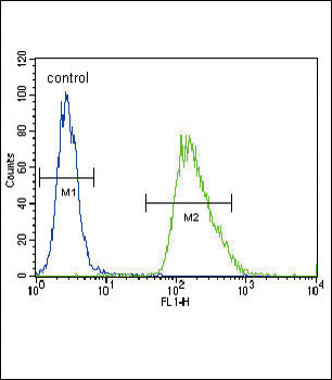
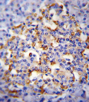
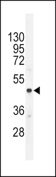
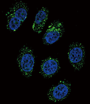
−| Tested Applications | FC, IF, IHC-P, WB |
|---|---|
| Dilution Range | IF: 1:10~50, WB: 1:1000, IHC-P: 1:10~50, FC: 1:10~50 |
| Reactivity | Human |
Key Properties
−| Antibody Type | Primary Antibody |
|---|---|
| Host | Rabbit |
| Clonality | Polyclonal |
| Isotype | Rabbit IgG |
| Clone No. | RB19424 |
| Target | This CMGA antibody is generated from rabbits immunized with a KLH conjugated synthetic peptide between 376-404 amino acids from the C-terminal region of human CMGA. |
| Molecular Weight | 50688 Da |
| Conjugation | Unconjugated |
Storage & Handling
−| Storage | Maintain refrigerated at 2-8°C for up to 2 weeks. For long term storage store at -20°C in small aliquots to prevent freeze-thaw cycles |
|---|---|
| Form/Appearance | Purified polyclonal antibody supplied in PBS with 0.09% (W/V) sodium azide. This antibody is purified through a protein A column, followed by peptide affinity purification. |
| Expiration Date | 12 months from date of receipt. |
| Disclaimer | For research use only |
Alternative Names
−Chromogranin-A, CgA, Pituitary secretory protein I, SP-I, Vasostatin-1, Vasostatin I, Vasostatin-2, Vasostatin II, EA-92, ES-43, Pancreastatin, SS-18, WA-8, WE-14, LF-19, AL-11, GV-19, GR-44, ER-37, CHGA
Similar Products
−- Item 1 of 4
CMGA Antibody (C-term) [orb1439009]
FC, IF, IHC-P, WB
Human
Rabbit
Polyclonal
Unconjugated
50 μl, 100 μl
Quality Guarantee
Explore bioreagents carefree to elevate your research. All our products are rigorously tested for performance. If a product does not perform as described on its datasheet, our scientific support team will provide expert troubleshooting, a prompt replacement, or a refund. For full details, please see our Terms & Conditions and Buying Guide. Contact us at [email protected].
Quick Database Links
Gene Symbol
This CMGA antibody is generated from rabbits immunized with a KLH conjugated synthetic peptide between 376-404 amino acids from the C-terminal region of human CMGA.
UniProt
RefSeq (Protein):NP_001266.1
UniProt Details
− No UniProt data available
NCBI Reference Sequences
−Associated Accession Numbers
Curated reference sequences for the gene transcript and protein product| Protein | NP_001266.1 |
|---|
Documents Download
Datasheet
Product Information
Request a Document
Protocol Information
WB
Western Blot (IB, immunoblot)
IHC-P
Immunohistochemistry Paraffin
FC
Flow Cytometry
IF
Immunofluorescence
CMGA Antibody (C-term) (orb1937956)

- 0.0
Based on 0 reviews
Participating in our Biorbyt product reviews program enables you to support fellow scientists by sharing your firsthand experience with our products.
Login to Submit a ReviewAvailable Sizes
Select a size below
Free Secondary Antibody (20 ul)0/0
Please add an antibody product to your cart first.