You have no items in your shopping cart.
Description
Research Area
Pharmacology & Drug Discovery
Images & Validation
−Item 1 of 3
| Tested Applications | ELISA, ICC, IF, IHC, WB |
|---|---|
| Dilution Range | WB (1:1000); ICC/IF (1:50); ELISA (1:1000); IHC: (1:25) optimal dilutions for assays should be determined by the user. |
| Reactivity | All |
| Application Notes |
Key Properties
−| Host | Mouse |
|---|---|
| Clonality | Monoclonal |
| Isotype | IgG1 |
| Clone No. | 6C2.1 |
| Immunogen | Synthetic L-Citrulline conjugated to Keyhole Limpet Hemocyanin (KLH). |
| Target | Citrulline |
| Purification | Protein G Purified |
| Conjugation | Biotin |
Storage & Handling
−| Storage | Conjugated antibodies should be stored according to the product label |
|---|---|
| Buffer/Preservatives | 136.36mM Ethanolamine, 133.23 mM Chlorides, 9.55mM Phosphates, 9.55mM Sodium Bicarbonate |
| Concentration | 1 mg/ml |
| Expiration Date | 12 months from date of receipt. |
| Disclaimer | For research use only |
Alternative Names
−L-Citrulline, 2-Amino-5-(carbamoylamino)pentanoic acid, Citrulline, Citrulline-modified protein, ACPA, Anti-citrullinated protein antibodies, Anti-citrullinated protein, Modified Citrulline, Citrulline-containing protein, Citrullinated protein
Similar Products
−- Item 1 of 1
Argininosuccinate synthetase 1, Biotinylated Antibody [orb341500]
ELISA, IF, IHC, IP, WB
Canine, Mouse, Rat
Bovine, Human
Goat
Polyclonal
Unconjugated
100 μg - Item 1 of 3

Citrulline-Histone H3 (Arg2/8/17) Recombinant Rabbit mAb, Biotin conjugated [orb2331409]
WB
Mouse, Rat
Human
Rabbit
Recombinant
Biotin
100 μlL-Citrulline Rabbit Polyclonal Antibody (Biotin) [orb456435]
ELISA, IF, IHC-Fr, IHC-P
All
Rabbit
Polyclonal
Biotin
100 μl


Quality Guarantee
Explore bioreagents carefree to elevate your research. All our products are rigorously tested for performance. If a product does not perform as described on its datasheet, our scientific support team will provide expert troubleshooting, a prompt replacement, or a refund. For full details, please see our Terms & Conditions and Buying Guide. Contact us at [email protected].

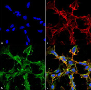
Immunocytochemistry/Immunofluorescence analysis using Mouse Anti-Citrulline Monoclonal Antibody, Clone 6C2.1. Tissue: Embryonic kidney epithelial cell line (HEK293). Species: Human. Fixation: 5% Formaldehyde for 5 min. Primary Antibody: Mouse Anti-Citrulline Monoclonal Antibody at 1:50 for 30-60 min at RT. Secondary Antibody: Goat Anti-Mouse Alexa Fluor 488 at 1:1500 for 30-60 min at RT. Counterstain: Phalloidin Alexa Fluor 633 F-Actin stain; DAPI (blue) nuclear stain at 1:250, 1:50000 for 30-60 min at RT. Magnification: 20X (2X Zoom). (A) DAPI (blue) nuclear stain. (B) Phalloidin Alexa Fluor 633 F-Actin stain. (C) Citrulline Antibody (D) Composite.
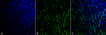
Immunohistochemistry analysis using Mouse Anti-Citrulline Monoclonal Antibody, Clone 6C2.1. Tissue: Colon tissue. Species: Mouse. Fixation: Formalin fixed, paraffin embedded. Primary Antibody: Mouse Anti-Citrulline Monoclonal Antibody at 1:25 for 1 hour at RT. Secondary Antibody: Goat Anti-Mouse IgG: Alexa Fluor 488. Counterstain: DAPI (blue) nuclear stain. Magnification: 63X. (A) DAPI (blue) nuclear stain. (B) Phalloidin Alexa Fluor 633 F-Actin stain. (C) O-GlcNAc Antibody (D) Composite.
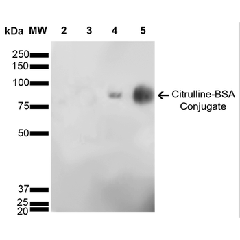
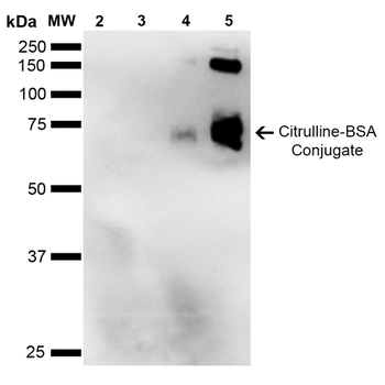
Western Blot analysis of Citrulline-BSA Conjugate showing detection of 67 kDa Citrulline protein using Mouse Anti-Citrulline Monoclonal Antibody, Clone 6C2.1. Lane 1: Molecular Weight Ladder (MW). Lane 2: BSA (0.5 μg). Lane 3: BSA (2.0 μg). Lane 4: Citrulline-BSA (0.5 μg). Lane 5: Citrulline-BSA (2.0 μg). Block: 5% Skim Milk in TBST. Primary Antibody: Mouse Anti-Citrulline Monoclonal Antibody at 1:1000 for 2 hours at RT. Secondary Antibody: Goat Anti-Mouse IgG: HRP at 1:2000 for 60 min at RT. Color Development: ECL solution for 5 min in RT. Predicted/Observed Size: 67 kDa.
Quick Database Links
Gene Symbol
Citrulline
Documents Download
Datasheet
Product Information
Request a Document
Protocol Information
WB
Western Blot (IB, immunoblot)
IHC
Immunohistochemistry
IF
Immunofluorescence
ICC
Immunocytochemistry
ELISA
Enzyme-linked Immunosorbent Assay (EIA)
Citrulline Antibody (Biotin) (orb396417)

- 0.0
Based on 0 reviews
Participating in our Biorbyt product reviews program enables you to support fellow scientists by sharing your firsthand experience with our products.
Login to Submit a ReviewAvailable Sizes
Select a size below
Choose Conjugation or Carrier Free Version
Free Secondary Antibody (20 ul)0/0
Please add an antibody product to your cart first.