You have no items in your shopping cart.
Featured
Description
Research Area
Epigenetics & Chromatin
Images & Validation
−Item 1 of 2
| Tested Applications | ICC, IF, WB |
|---|---|
| Dilution Range | WB (1:1000), ICC/IF (1:100) |
| Reactivity | Human |
| Application Notes |
Key Properties
−| Host | Mouse |
|---|---|
| Clonality | Monoclonal |
| Isotype | IgG1 |
| Clone No. | 5A7-2E11 |
| Immunogen | Synthetic peptide corresponding to a portion of human CENP-A |
| Target | CENPA |
| Molecular Weight | 18kDa |
| Purification | Protein G Purified |
| Conjugation | PerCP |
Storage & Handling
−| Storage | Conjugated antibodies should be stored according to the product label |
|---|---|
| Buffer/Preservatives | 95.46mM Phosphate, 2.48mM MES and 2mM EDTA |
| Concentration | 1 mg/ml |
| Expiration Date | 12 months from date of receipt. |
| Disclaimer | For research use only |
Alternative Names
−CENPA, CENP-A, Centromere autoantigen A, Histone H3-like centromeric protein A
Quality Guarantee
Explore bioreagents carefree to elevate your research. All our products are rigorously tested for performance. If a product does not perform as described on its datasheet, our scientific support team will provide expert troubleshooting, a prompt replacement, or a refund. For full details, please see our Terms & Conditions and Buying Guide. Contact us at [email protected].
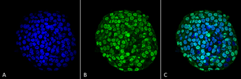
Immunocytochemistry/Immunofluorescence analysis using Mouse Anti-CENP-A Monoclonal Antibody, Clone 5A7-2E11. Tissue: Colon cancer cell line (HT-29). Species: Human. Fixation: 4% Formaldehyde for 15 min at RT. Primary Antibody: Mouse Anti-CENP-A Monoclonal Antibody at 1:100 for 60 min at RT. Secondary Antibody: Goat Anti-Mouse ATTO 488 at 1:100 for 60 min at RT. Counterstain: DAPI (blue) nuclear stain at 1:5000 for 5 min RT. Localization: Nucleus. Magnification: 60X.
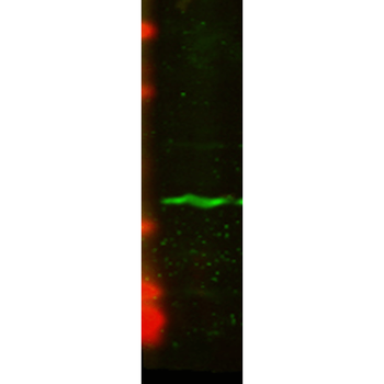
Western Blot analysis of Human U2OS cell lysate showing detection of CENP-A protein using Mouse Anti-CENP-A Monoclonal Antibody, Clone 5A7-2E11. Primary Antibody: Mouse Anti-CENP-A Monoclonal Antibody at 1:1000.
Quick Database Links
UniProt Details
− No UniProt data available
NCBI Gene Details
− No NCBI Gene data available
NCBI Reference Sequences
−Associated Accession Numbers
Curated reference sequences for the gene transcript and protein product| Protein | NP_001035891.1 |
|---|
Documents Download
Datasheet
Product Information
Request a Document
Protocol Information
WB
Western Blot (IB, immunoblot)
IF
Immunofluorescence
ICC
Immunocytochemistry
CENPA Antibody (PerCP) (orb148114)

- 0.0
Based on 0 reviews
Participating in our Biorbyt product reviews program enables you to support fellow scientists by sharing your firsthand experience with our products.
Login to Submit a ReviewAvailable Sizes
Select a size below
Choose Conjugation or Carrier Free Version
Free Secondary Antibody (20 ul)0/0
Please add an antibody product to your cart first.