You have no items in your shopping cart.
Description
Research Area
Cell Biology
Images & Validation
−Item 1 of 5
| Tested Applications | IF, IHC-P, WB |
|---|---|
| Dilution Range | IF - 1:100, WB - 1:1000, IHC-P - 1:100 |
| Reactivity | Human, Mouse |
| Predicted Reactivity | Zebrafish |
Key Properties
−| Host | Rabbit |
|---|---|
| Clonality | Polyclonal |
| Isotype | Rabbit IgG |
| Immunogen | This Cellular Apoptosis Susceptibility antibody is generated from rabbits immunized with a KLH conjugated synthetic peptide between 55-84 amino acids from the N-terminal region of human Cellular Apoptosis Susceptibility. Antigen Region: 55-84 aa. |
| Target | CSE1L |
| Molecular Weight | 110417 Da |
| Conjugation | Unconjugated |
Storage & Handling
−| Storage | Maintain refrigerated at 2-8°C for up to 2 weeks. For long term storage store at -20°C in small aliquots to prevent freeze-thaw cycles |
|---|---|
| Form/Appearance | Purified polyclonal antibody supplied in PBS with 0.09% (W/V) sodium azide. This antibody is prepared by Saturated Ammonium Sulfate (SAS) precipitation followed by dialysis against PBS. |
| Expiration Date | 12 months from date of receipt. |
| Disclaimer | For research use only |
Alternative Names
−Exportin-2, Exp2, Cellular apoptosis susceptibility protein, Chromosome segregation 1-like protein, Importin-alpha re-exporter, CSE1L, CAS, XPO2
Quality Guarantee
Explore bioreagents carefree to elevate your research. All our products are rigorously tested for performance. If a product does not perform as described on its datasheet, our scientific support team will provide expert troubleshooting, a prompt replacement, or a refund. For full details, please see our Terms & Conditions and Buying Guide. Contact us at [email protected].
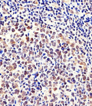
Immunohistochemical analysis of paraffin-embedded H. tonsil section using CSE1L Antibody. Diluted at 1:100 dilution. A peroxidase-conjugated goat anti-rabbit IgG at 1:400 dilution was used as the secondary antibody, followed by DAB staining.
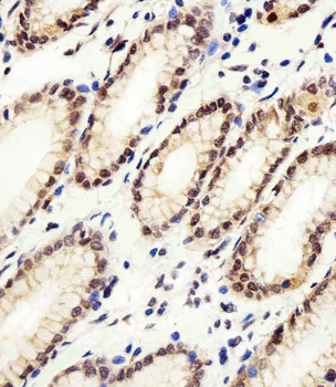
Immunohistochemical analysis of paraffin-embedded H. stomach section using CSE1L Antibody. Diluted at 1:100 dilution. A peroxidase-conjugated goat anti-rabbit IgG at 1:400 dilution was used as the secondary antibody, followed by DAB staining.
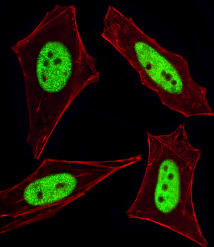
Fluorescent image of HeLa cells stained with Cellular Apoptosis Susceptibility Antibody (N-term). Diluted at 1:100 dilution. An Alexa Fluor 488-conjugated goat anti-rabbit lgG at 1:400 dilution was used as the secondary antibody (green). Cytoplasmic actin was counterstained with Alexa Fluor 555 conjugated with Phalloidin (red).
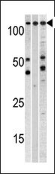
Western blot analysis of anti-CSE1L Pab in, from left to right, A375, CEM, and mouse heart cell line lysates (35 ug/lane). CSE1L (arrow) was detected using the purified Pab.
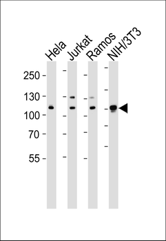
Western blot analysis of lysates from Hela, Jurkat, Ramos, mouse NIH/3T3 cell line (from left to right), using CSE1L Antibody. Diluted at 1:1000 at each lane. A goat anti-rabbit IgG H&L (HRP) at 1:5000 dilution was used as the secondary antibody. Lysates at 35 ug per lane.
Quick Database Links
UniProt Details
− No UniProt data available
NCBI Reference Sequences
−Associated Accession Numbers
Curated reference sequences for the gene transcript and protein product| Protein | NP_001307.2, NP_001243064.1 |
|---|
Documents Download
Datasheet
Product Information
Request a Document
Protocol Information
WB
Western Blot (IB, immunoblot)
IHC-P
Immunohistochemistry Paraffin
IF
Immunofluorescence
Cellular Apoptosis Susceptibility Antibody (C-term) (orb1932372)

- 0.0
Based on 0 reviews
Participating in our Biorbyt product reviews program enables you to support fellow scientists by sharing your firsthand experience with our products.
Login to Submit a ReviewAvailable Sizes
Select a size below
Choose Conjugation or Carrier Free Version
Free Secondary Antibody (20 ul)0/0
Please add an antibody product to your cart first.