
You have no items in your shopping cart.
Description
Research Area
Epigenetics
Images & Validation
−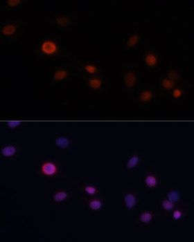
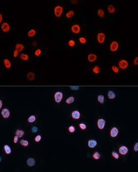
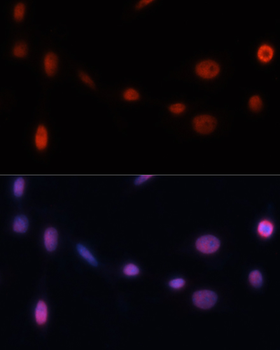
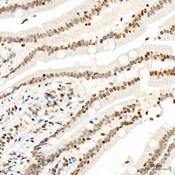
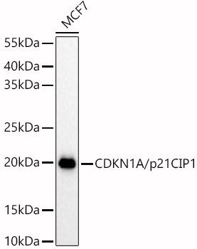
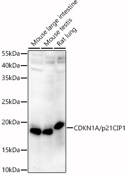
Item 1 of 6
| Tested Applications | ELISA, IF, IHC-P, WB |
|---|---|
| Dilution Range | WB: 1:500-2000, IHC-P: 1:50-200, IF/ICC: 1:20-100 |
| Reactivity | Human, Mouse, Rat |
Key Properties
−| Host | Rabbit |
|---|---|
| Clonality | Polyclonal |
| Isotype | IgG |
| Immunogen | A synthetic peptide corresponding to a sequence within amino acids 1-164 of human CDKN1A/p21CIP1 (NP_000380.1). |
| Molecular Weight | 18kDa |
| Purification | Affinity purification |
| Conjugation | Unconjugated |
Storage & Handling
−| Storage | Maintain refrigerated at 2-8°C for up to 2 weeks. For long term storage store at -20°C in small aliquots to prevent freeze-thaw cycles. |
|---|---|
| Buffer/Preservatives | PBS with 0.09% Sodium azide,50% glycerol,pH7.3. |
| Concentration | 1 mg/ml |
| Expiration Date | 12 months from date of receipt. |
| Disclaimer | For research use only |
Alternative Names
−anti CAP20 antibody, anti CDKN1 antibody, anti CIP1 antibody, anti MDA-6 antibody, anti P21 antibody, anti SDI1 antibody, anti WAF1 antibody, anti p21CIP1 antibody

Quality Guarantee
Explore bioreagents carefree to elevate your research. All our products are rigorously tested for performance. If a product does not perform as described on its datasheet, our scientific support team will provide expert troubleshooting, a prompt replacement, or a refund. For full details, please see our Terms & Conditions and Buying Guide. Contact us at [email protected].
Quick Database Links
UniProt
UniProt Details
− No UniProt data available
Protocol Information
WB
Western Blot (IB, immunoblot)
IHC-P
Immunohistochemistry Paraffin
IF
Immunofluorescence
ELISA
Enzyme-linked Immunosorbent Assay (EIA)
Filter by Applications
Filter by Species
Kimoto, Saki et al. Nacre extract derived from Pinctada fucata mitigates skeletal muscle aging by suppressing senescence-associated inflammation and preserving contractile features in naturally aged mice and C2C12 myotubes Asian Pacific Journal of Tropical Biomedicine, (2026)
Applications
IF
Reactivity
Mouse
Chutikorn Nopparat, Varunya Chantadul, Kannika Permpoonputtana, Piyarat Govitrapong The anti-inflammatory effect of melatonin in SH-SY5Y neuroblastoma cells exposed to sublethal dose of hydrogen peroxide Mechanisms of Ageing and Development, 164, 49 (2017)
Applications
WB
Reactivity
Human
Momoko Kawaminami et al. Nacre extract attenuates age-related functional and tissue alterations under post-onset intervention conditions in two murine aging models Biogerontology, 27, 114 (2026) (2026)
Applications
IF, IHC
Reactivity
Mouse